9, 7, 2014

What is the Femoral Vein?

2022-01-03T13:29:29-08:00

The femoral vein is a large blood vessel of the leg that allows deoxygenated blood to travel to the heart and lungs to become oxygenated. It is located deep within the muscles of the thigh beginning just above the knee (at the adductor canal it is the continuation of the popliteal vein) and ends at the groin level (specifically, it ends at the inferior margin of the inguinal ligament where it becomes the external iliac vein.  It accompanies the femoral artery in the femoral sheath.  In this ultrasound image from La Jolla Vein Care, notice that the femoral vein runs along the same course as the femoral artery, which provides oxygenated blood from the heart and lungs to the rest of the body. The arteries and veins carry blood in opposite directions.  Ultrasound imaging detects the direction of blood flow and in this image, it femoral vein is ‘blue’ depicting blood flow moving toward the heart and the femoral artery is ‘red’ demonstrating blood flow away from the heart.

Ultra Sound detects Femoral Vein

Ultrasound image of the femoral vein and femoral artery. Ultrasound imaging detects the direction of blood flow and in this La Jolla Vein Care image, the femoral vein is ‘blue’ depicting blood flow moving toward the heart and the femoral artery is ‘red’ demonstrating blood flow away from the heart. Notice that the femoral vein and artery are located within the muscle. For orientation purposes, the skin is located at the top of the image.


What is the Femoral Vein?2022-01-03T13:29:29-08:00

Foam Treatment of Venous Malformations

2021-11-05T13:22:04-07:00
DSCF0086 copy

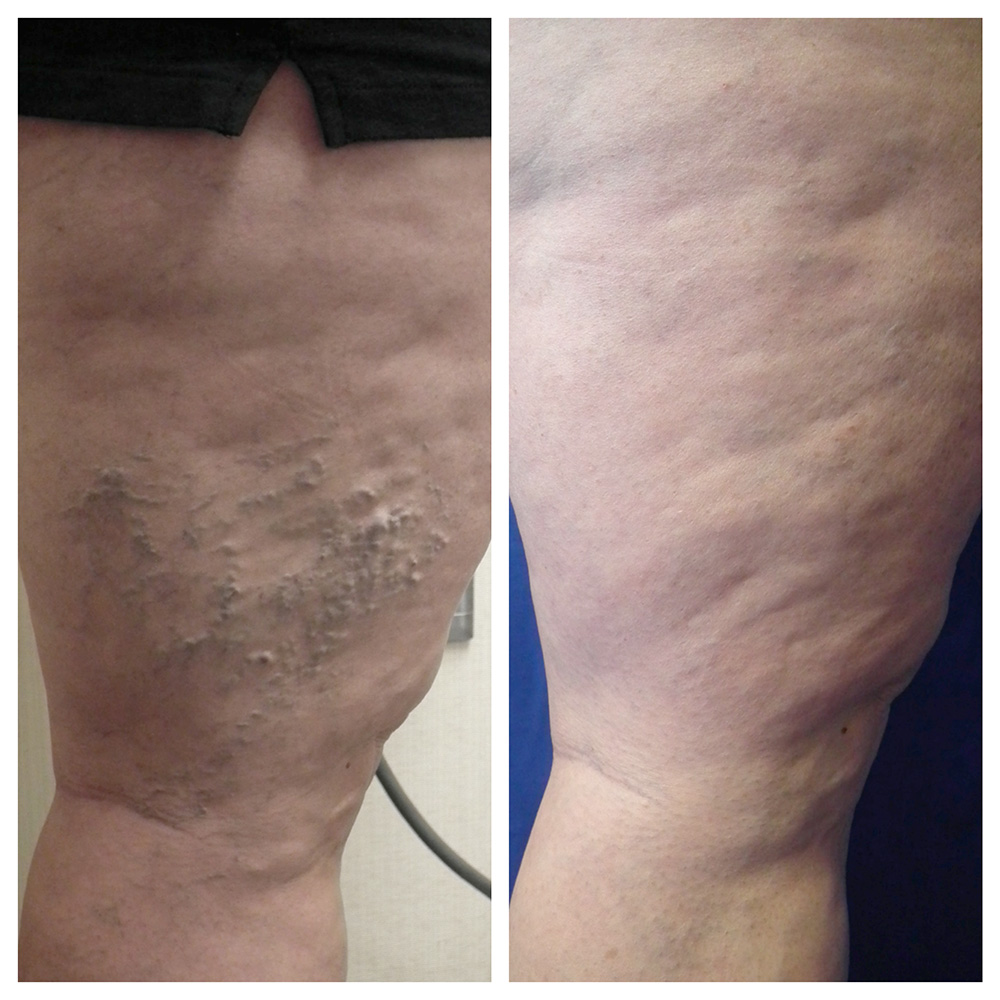
Venous malformation of the lateral leg before treatment. Venous malformations appear like abnormally dilated bluish vessels near the surface of the skin. They appear in clusters and look different than typical varicose veins. They often appear during childhood, as opposed to varicose veins.

Venous malformations comprise either superficial or deep veins that are abnormally formed and dilated.  Although they usually are present at birth, they may not be seen until years later into adolescence or even adulthood. The natural history of a venous malformation is slow, steady enlargement. However, events such as surgery, trauma, infection, or hormonal changes associated with puberty, pregnancy or menopause may cause rapid expansion.

At La Jolla Vein Care, we frequently evaluate and treat superficial venous malformations of the legs. Prior to treatment, it is important to have thorough diagnostic imaging, such as an ultrasound examination to ensure an accurate diagnosis of a pure venous malformation.  If an arterial connection (arterial venous malformation) is suspected on ultrasound evaluation, further imaging with CT or MR arteriogram may be necessary. Venous malformations can also occur in other syndromes, such as Klippel -Trenaunay syndrome and may involve the abnormal development of the deep veins.

Pure venous malformations can be treated without surgery. Foam sclerotherapy is a common treatment type.  Foam sclerotherapy uses a technique to inject a foamed sclerosing solution into the cluster of varicose veins, which will cause them to collapse and eventually dissolve.

DSCF1899

Venous malformation after treatment with ultrasound-guided foam sclerotherapy at La Jolla Vein Care. Treatment can reduce the symptoms associated with venous malformations such as leg pain, aching and throbbing.

Foam Treatment of Venous Malformations2021-11-05T13:22:04-07:00

8 Warning Signs of Vein Disease: #4 Swollen Ankles

2021-11-05T04:26:19-07:00
After

Ankle swelling related to varicose veins and venous insufficiency may be subtle, leaving indentations from socks.

Swollen Ankles at Night

Thick, swollen ankles are signs that blood or other fluid is congested in the leg and/or leg veins. Over time, damaged vein walls can become even more stretched out and permeable, allowing fluid and protein to filter from the veins into surrounding leg tissue. When you lie down at night, the pressure from gravity is equalized across your leg. Usually, vein related swelling (venous edema) becomes apparent later in the day or worse throughout the day and improved with leg elevation or overnight during sleep.  Often in the morning, there may be no swelling. But, as the day progresses gravity causes poo

Pooling of blood around the ankle in incompetent veins. Venous insufficiency is one of the most common causes of ankle swelling that worsens throughout the day. Sometimes, the swelling can be subtle, leaving indentations from sock lines. Or, it may cause the skin to feel firm, shiny, or puffy by the end of the day.

If you have swelling, you should discuss it with your healthcare provider. Venous insufficiency can be diagnosed with a venous duplex sonogram, which is a non-invasive study of the leg veins.

8 Warning Signs of Vein Disease: #4 Swollen Ankles2021-11-05T04:26:19-07:00

8 Warning Signs of Vein Disease: #3 Open Sores or Ulcers on the Lower Leg

2014-07-02T00:16:13-07:00
Before

Venous leg ulcers are the result of chronic venous insufficiency and venous hypertension, which causes the skin to break down.

When chronic venous insufficiency reaches its most serious point, ulcers may appear on the lower leg. These open ulcers are the result of blood leaking into the leg tissue and damaging the skin. Open sores need to be treated by a doctor immediately.

8 Warning Signs of Vein Disease: #3 Open Sores or Ulcers on the Lower Leg2014-07-02T00:16:13-07:00
Go to Top