25, 8, 2023

Saphenous Venous Reflux

2023-05-24T16:48:10-07:00

ClosureFast™ an endovenous radiofrequency ablation (RFA) procedure for saphenous vein reflux,

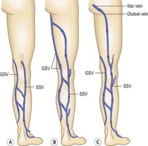
for the backward flow of blood (or “Venous reflux”) in your saphenous vein(s). The great saphenous veins and small saphenous veins are the two main superficial veins of the leg. They run along the inner leg and the back of the leg, respectively.

This minimally invasive procedure can be performed in the office in less than an hour and patients usually return to their usual level of activity the same day.

HOW DOES THE TREATMENT WORK?

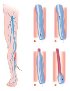
The skin is numbed with lidocaine, then a tiny wire and the Closurefast® catheter are inserted into the vein.  The catheter delivers radio-frequency energy to the vein wall, causing it to seal shut.  The remaining healthy veins continue to bring blood back to the heart.

WHAT SHOULD I EXPECT ON THE DAY OF TREATMENT?

The procedure is performed with local anesthesia, but many patients elect to use a mild oral sedative (Valium), which is taken after checking in and
saphenous

Diagram of endovenous laser ablation (EVLA) procedure

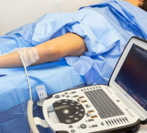
completing all paperwork.  You will change into a gown and leave underwear on.  Depending on the vein to be treated, you will lay on your back or on your belly.  We do our best to make special accommodations (for example, if you cannot lie flat or cannot bend a knee very well) with body positioning and using pillows.  We will do our best to make you comfortable.  Then, we will give you the option of watching a movie on Netflix or listening to music.  Once you are comfortable, your leg (s) will be prepped with a cleansing solution for the sterile procedure.  The doctor will perform an ultrasound to map the vein (s) to be treated.  Then, a numbing agent (lidocaine) will be injected into the skin. In the numb area of the skin, a tiny puncture is made to pass the radiofrequency catheter.  Your doctor will then use a needle to administer a combination of cool saline and local anesthetic around the vein either in the thigh or calf (depending on which vein is treated).  This solution numbs the vein and insulates it from the surrounding tissue.  After the numbing solution is applied, the vein is painlessly treated with radiofrequency energy.

Once your vein has been treated, we will clean your leg and apply a compression stocking which you will wear for 72 hours continuously.  You will walk for 30 minutes prior to getting in your car.


“Bringing Experts Together for Unparalleled Vein and Vascular Care”

La Jolla Vein & Vascular (formerly La Jolla Vein Care) is committed to bringing experts together for unparalleled vein and vascular care. 

Nisha Bunke, MD, Sarah Lucas, MD, and Elliot DeYoung, MD are specialists who combine their experience and expertise to offer world-class vascular care. 

Our accredited center is also a nationally known teaching site and center of excellence. 

For more information on treatments and to book a consultation, please give our office a call at 858-550-0330. 

 For a deeper dive into vein and vascular care, please check out our Youtube Channel at this link, and our website https://ljvascular.com

For more information on varicose veins and eliminating underlying venous insufficiency,

Please follow our social media Instagram Profile and Tik Tok Profile for more fun videos and educational information. 

For more blogs and educational content, please check out our clinic’s blog posts


Saphenous Venous Reflux2023-05-24T16:48:10-07:00

18, 4, 2023

Saphenous Vein Reflux Treatment

2023-02-15T15:47:21-08:00

ClosureFast™ an endovenous radiofrequency ablation (RFA) procedure

for the backward flow of blood (or “Venous reflux”) in your saphenous vein(s). The great saphenous veins and small saphenous veins are the two main superficial veins of the leg. They run along the inner leg and the back of the leg, respectively.

This minimally invasive procedure can be performed in the office in less than an hour and patients usually return to their usual level of activity the same day.

saphenous

HOW DOES THE TREATMENT WORK?

The skin is numbed with lidocaine, then a tiny wire and the Closurefast® catheter are inserted into the vein.  The catheter delivers radio-frequency energy to the vein wall, causing it to seal shut.  The remaining healthy veins continue to bring blood back to the heart.

WHAT SHOULD I EXPECT ON THE DAY OF TREATMENT?

The procedure is performed with local anesthesia, but many patients elect to use a mild oral sedative (Valium), which is taken after checking in and

Diagram of endovenous laser ablation (EVLA) procedure

completing all paperwork.  You will change into a gown and leave underwear on.  Depending on the vein to be treated, you will lay on your back or on your belly.  We do our best to make special accommodations (for example, if you cannot lie flat or cannot bend a knee very well) with body positioning and using pillows.  We will do our best to make you comfortable.  Then, we will give you the option of watching a movie on Netflix or listen to music.  Once you are comfortable, you leg (s) will be prepped with cleansing solution for the sterile procedure.  The doctor will perform an ultrasound to map the vein (s) to be treated.  Then, a numbing agent (lidocaine) will be injected into the skin. In the numb area of the skin, a tiny puncture is made to pass the radiofrequency catheter.  Your doctor will then use a needle to administer a combination of cool saline and local anesthetic around the vein either in the thigh or calf (depending on which vein is treated).  This solution numbs the vein and insulates it from the surrounding tissue.  After the numbing solution is applied, the vein is painlessly treated with radiofrequency energy.

Once your vein has been treated, we will clean your leg and apply a compression stocking which you will wear for 72 hours continuously.  You will walk for 30 minutes prior to getting in your car.


“Bringing Experts Together for Unparalleled Vein and Vascular Care”

La Jolla Vein & Vascular (formerly La Jolla Vein Care) is committed to bringing experts together for unparalleled vein and vascular care. 

Nisha Bunke, MD, Sarah Lucas, MD, and Elliot DeYoung, MD are specialists who combine their experience and expertise to offer world-class vascular care. 

Our accredited center is also a nationally known teaching site and center of excellence. 

For more information on treatments and to book a consultation, please give our office a call at 858-550-0330. 


For a deeper dive into vein and vascular care, please check out our Youtube Channel at this link.


For more information on varicose veins and eliminating underlying venous insufficiency, check this link out full of resources. 

Please follow our social media Instagram Profile and Tik Tok Profile for more fun videos and educational information. 


Saphenous Vein Reflux Treatment2023-02-15T15:47:21-08:00

26, 6, 2020

Understanding What Your Doctor Is Saying About Your Veins

2020-09-17T18:42:20-07:00

If you’re not a vascular specialist, it may be difficult to understand the words used to describe your vein condition. Vein terms are not used in everyday life.  Especially, when it comes to the anatomy of the veins. Below is a glossary of the terms, we as vein specialists, use on a daily basis but may be foreign to the patient.  This is provided from the American Vein and Lymphatic Society (AVLS) healthyveins.org.

Anatomical Vein Terms

Anterior: The front of a body part.

Posterior: Back of a body part.

Deep Leg Vein: The veins that are deep in the thigh and leg that carry blood back to the heart.

Connective Tissue: This strong tissue is the white tissue that covers muscle.

Great Saphenous Vein: (sometimes called GSV or long saphenous vein) A long vein that can be seen just in front of the anklebone. This vein travels along the inside of the leg and thigh (about one-half inch beneath the skin in the thigh) until it empties into the deep vein called the common femoral vein in the groin.

Perforating Vein: A vein that passes directly from a superficial vein to a deep vein.

Popliteal Vein: The deep vein located behind the knee. The small saphenous vein originates from the popliteal vein.

Small Saphenous Vein: (sometimes called SSV, Lesser Saphenous Vein, or LSV) A superficial vein that starts at the outside of the foot and travels up the back of the calf where it empties into the deep vein (popliteal vein) in the crease of the knee.

Spider Vein: A tiny varicose vein that may be blue or red color that does not protrude above the skin surface and frequently looks like a spider. Common medical terms you may hear for spider veins are telangiectasias and telangiectatic veins. Slightly larger varicose veins that do not protrude above the skin are called reticular veins.

Superficial Vein: Any vein in the lower extremity above the deep fascia that covers the muscles of the thigh and leg.

Varicose Vein: A bulging vein that protrudes past the skin surface and usually measures greater than one-fourth of an inch (6.4 mm) in diameter.

Vein Function Terms

Competent Vein: Blood flow occurs in the proper direction back to the heart. Also referred to as Having No Reflux Or Normal Flow Direction.

Incompetent Vein: Blood flows in the wrong direction. Also referred to as a vein that has reflux.

Reflux: Blood that flows backward in the veins.


Anatomical Images and Explanations

Normal Blood Flow: After the blood has been replenished with oxygen in the lungs, it is pumped to the body by the heart. Blood that is pumped to the lower extremities is pumped back to the heart partially by the action of the calf muscle pump. The blood returning from the lower extremities in the deep and superficial veins goes past a series of one-way valves. These valves stop the blood from flowing backwards in the veins.

Reflux: The valves in the veins close just as blood begins to flow backwards. If the valves do not close properly, the blood falls backwards through the poorly closing or leaking valves. The veins downstream that are now unprotected by valves further upstream are exposed to the weight of an increasingly high column of blood. These downstream veins cannot endure the pressure of the column of blood and expand becoming snake-like in appearance. This causes the veins to bulge through the skin surface and become varicose veins.

Understanding What Your Doctor Is Saying About Your Veins2020-09-17T18:42:20-07:00

9, 10, 2015

What’s So Great About the Great Saphenous Vein?

2022-04-24T23:30:58-07:00

Great Saphenous vein and short saphenous vein

The great saphenous vein (GSV), previously referred to as the long saphenous vein, is a superficial leg vein that runs from the top of the thigh near the groin, down the inner thigh all the way to the inner ankle. The top blue arrow in this diagram points to the location of the great saphenous vein.

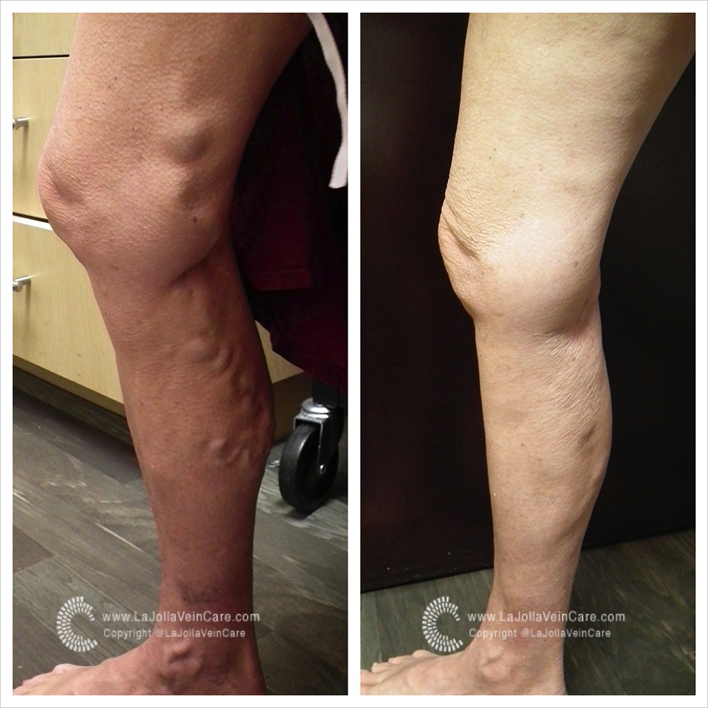
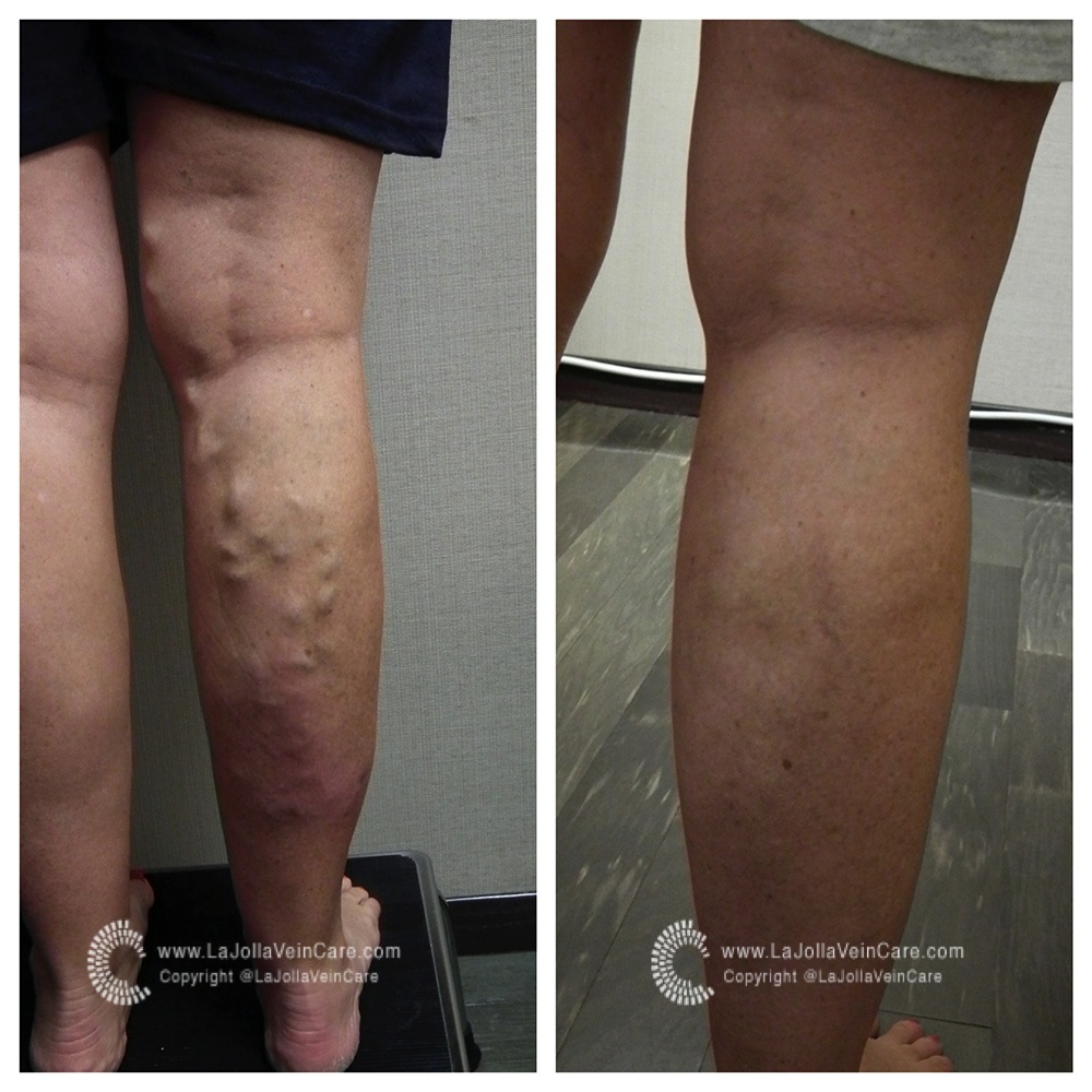
The Great Saphenous vein is responsible for varicose veins about 80% of the time. When varicose veins appear in the inner thigh or calf areas, the GSV is often the culprit (see picture). The other superficial vein is largely responsible for varicose veins in the small saphenous vein. The small saphenous vein (SSV) runs along the back of the calf. The SSV was previously referred to as the short saphenous vein. Varicose veins on the backside of the leg are often caused by leaky valves within the small saphenous vein.

varicose-DSCF5885_fotorvaricose-21after_fotor


What’s So Great About the Great Saphenous Vein?2022-04-24T23:30:58-07:00

15, 5, 2014

What If I need my Great Saphenous Vein for Bypass Someday?

2021-11-04T16:18:45-07:00
GSV1

The Great Saphenous Vein is a vein of the superficial venous system that drains blood from the skin and tissues into the deep system. It is most commonly the root of the problem in people with varicose veins. When the great saphenous vein is dysfunctional, removing it is the first step of vein treatment.

A great saphenous vein that needs treatment is not functioning properly. When a vein is diseased, the walls become weak and the vessel can dilate or grow triple its’ normal size. This is not the type of vessel that would be ideal to place in a diseased heart. While the saphenous vein is a common vein that is harvested for bypass grafts, there are other options such as the internal thoracic artery in the chest and the radial artery which is in the arm. With these options, an artery replaces an artery and the removal of the valves inside the veins is not necessary. It is important for the entire circulatory system to be functioning properly, together.

What If I need my Great Saphenous Vein for Bypass Someday?2021-11-04T16:18:45-07:00

25, 3, 2014

What is a Venous Insufficiency / Duplex Ultrasound Test?

2021-11-04T00:54:25-07:00

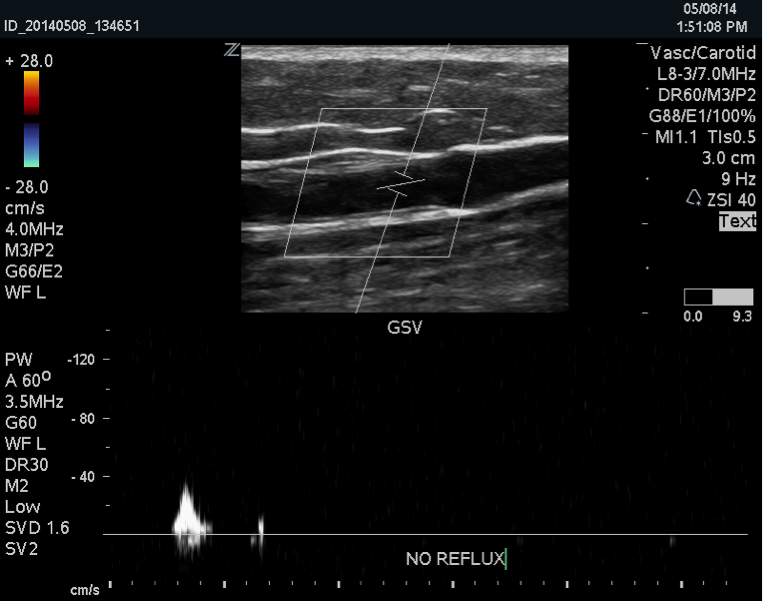
The Duplex Ultrasound examination, also called a venous insufficiency study allows us to visualize the blood vessels that are not visible to the naked eye, even blood vessels that are deep within the muscles.  The ultrasound examination is used to both identify the veins that have faulty valves and to map the anatomy of the veins, creating a ‘road map.’  This is necessary to make an accurate assessment of the cause and extent of the varicose veins, as well as to formulate the best treatment plan.  This should be done for any individual being evaluated for varicose veins, leg swelling, patients who have failed prior treatment, patients who are symptomatic and in some patients with certain anatomic patterns of spider veins.

Duplex ultrasound uses sound waves to get images of your blood vessels.  It also helps determine how fast blood moves through the vessels.

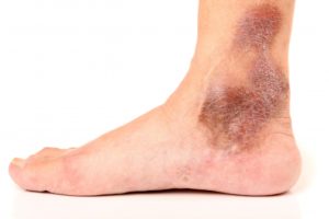
Darkening of skin, and itchiness are a sign of chronic venous insufficiency or venous reflux disease

Darkening of skin, and itchiness are a sign of chronic venous insufficiency or venous reflux disease

At La Jolla Vein Care, the venous duplex study will be performed by one of our registered vascular technologists, who have specialized training in the science of vascular sonography (RVT). It is performed in the standing position, which is important in obtaining an accurate study result.  This examination takes approximately 45-60 minutes. Following the study, the doctor will personally discuss the results with our patients to ensure a complete understanding of the ultrasound findings.  Insurance typically covers this type of diagnostic study.


What is a Venous Insufficiency / Duplex Ultrasound Test?2021-11-04T00:54:25-07:00
Go to Top