Unveiling the Road to Better Health: The Power of Carotid Artery Ultrasound
LJVascular2023-10-11T17:22:03-07:00Unveiling the Road to Better Health: The Power of Carotid Artery Ultrasound
Envision having the ability to peer into the intricate passageways that serve as vital conduits for blood traveling to your brain, allowing you to detect potential threats before they strike. Carotid Artery Ultrasounds provide precisely this capability, offering a non-invasive window into your carotid arteries to fortify the defense against strokes. In this blog post, we shall embark on a journey into the realm of carotid artery ultrasounds, elucidating their critical role in stroke prevention and acquainting you with the risk factors that demand your attention.
Unveiling the Significance of Carotid Artery Ultrasounds
Carotid Artery Ultrasounds are more than mere routine assessments; they represent powerful weapons in the battle against strokes. These ultrasounds serve as vital instruments for appraising the condition of the carotid arteries, responsible for supplying the brain with oxygen-rich blood. Their primary mission is to unearth potential issues that may pave the way for strokes, particularly in individuals who have manifested symptoms of a Transient Ischemic Attack (TIA) or are perched atop the precipice of stroke risk.
Deciphering the Transient Ischemic Attack (TIA)
Before we immerse ourselves in the intricacies of carotid artery ultrasounds, it’s essential to shed light on Transient Ischemic Attacks, commonly known as TIAs. These episodes serve as warnings that the brain’s blood supply faces a momentary interruption. The symptoms associated with TIAs are akin to those of a stroke and may encompass:
- Sudden numbness in the face, arm, or leg, especially on one side of the body.
- Difficulty speaking or slurred speech.
- Vision anomalies, such as abrupt blurred or double vision.
- Dizziness or loss of balance.
- An intense headache without any apparent cause.
TIAs mirror the symptoms of a stroke, but the fundamental distinction lies in their transient nature. TIAs are ephemeral episodes, typically lasting only a few minutes to a couple of hours, and they do not inflict permanent damage. However, they act as substantial red flags, signaling the potential for a forthcoming stroke.
Assessing Stroke Risk through Ultrasound Screenings
Carotid artery ultrasounds prove instrumental in the assessment of stroke risk, particularly in individuals who have encountered TIA symptoms. Here is how these screenings operate:
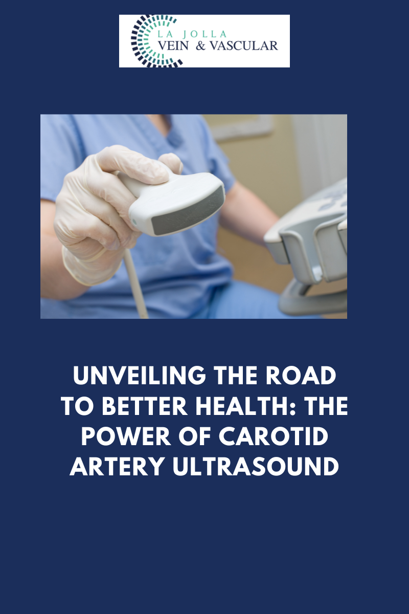
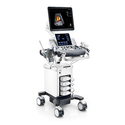
- Non-Invasive Imaging: Carotid artery ultrasounds harness non-invasive imaging technology to render the carotid arteries in the neck visible. By using high-frequency sound waves, these ultrasounds craft detailed images of these pivotal blood vessels.
- Discerning Blockages: The ultrasound aids in the detection of blockages or the buildup of plaque within the carotid arteries. These impediments can diminish the blood flow to the brain, amplifying the vulnerability to a stroke.
- Early Intervention: The early detection of blockages empowers healthcare providers to take preemptive actions aimed at stroke prevention. This may encompass the administration of medications or surgical procedures designed to clear the arteries.
Familiarize Yourself with the Risk Factors for Strokes and Arterial Disease
Gaining a comprehensive understanding of the risk factors associated with strokes and arterial disease represents the initial stride on the path to prevention. The following risk factors warrant your attention:
- High Blood Pressure: Hypertension reigns as a principal contributor to stroke, underscoring the imperative nature of blood pressure management.
- Tobacco Usage: Smoking escalates the risk of arterial disease and strokes considerably. The act of quitting smoking ranks among the most effective measures for curbing this risk.
- Diabetes: Individuals contending with diabetes encounter an elevated risk of strokes. Prudent management of blood sugar levels assumes paramount significance.
- Family History: A familial history intertwined with strokes or arterial disease can elevate your risk.
- Age: The risk of a stroke surges with age, particularly following the age of 55.
- Obesity: Excessive weight may contribute to heightened blood pressure and various other risk factors.
- Sleep Apnea: Untreated sleep apnea can elevate the risk of stroke due to interrupted breathing during sleep.
Carotid Artery Ultrasounds transcend their identity as mere medical procedures; they stand as vigilant protectors of your cerebral well-being. If you have encountered TIA symptoms or belong to the categories at increased risk, the discussion of carotid artery ultrasounds with your healthcare provider becomes a proactive step toward stroke prevention. Through the comprehension of warning signs and risk factors, you retain the power to command your vascular health and reduce the likelihood of a stroke rewriting the narrative of your life.
“Bringing Experts Together for Unparalleled Vein and Vascular Care”
La Jolla Vein & Vascular (formerly La Jolla Vein Care) is committed to bringing experts together for unparalleled vein and vascular care.
Nisha Bunke, MD, Sarah Lucas, MD, and Amanda Steinberger, MD are specialists who combine their experience and expertise to offer world-class vascular care.
Our accredited center is also a nationally known teaching site and center of excellence.
For more information on treatments and to book a consultation, please give our office a call at 858-550-0330.
For a deeper dive into vein and vascular care, please check out our Youtube Channel at this link, and our website https://ljvascular.com
For more information on varicose veins and eliminating underlying venous insufficiency,
Please follow our social media Instagram Profile and Tik Tok Profile for more fun videos and educational information.
For more blogs and educational content, please check out our clinic’s blog posts!


