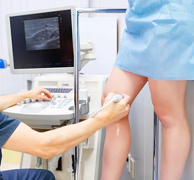
What Leg Venous Vein Ultrasound Can Uncover About Your Veins

2021-12-31T13:46:54-08:00

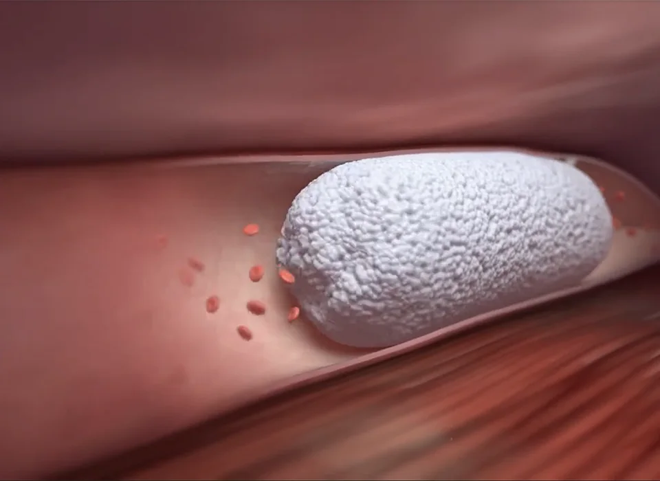
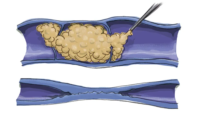
Most vein disease is not visible to the naked eye.

What Leg Vein Ultrasound Can Uncover About Your Veins: blood clots and leaky valves

Leg Vein Ultrasound

We can see beneath the surface of the skin with ultrasound. Duplex ultrasound combines Doppler flow information and conventional imaging information, sometimes called B-mode, to allow physicians to see the structure of your blood vessels. Duplex ultrasound uses sound waves to get images of your blood vessels.  It also helps determine how fast blood moves through the vessels. It can also be useful to estimate the diameter of a blood vessel as well as the amount of obstruction, if any, in the blood vessel. Using duplex ultrasound technology, the structure of your blood vessels, the valve function, the movement of your red blood cells, and the direction of blood flow through the vessels, any blockages, blood clots, and leaky valves can be seen.

Venous Reflux (leaky valves)

Duplex ultrasound of the leg veins

Ultrasounds of the leg veins that look for leaky valves (venous reflux) are usually performed in the standing position.

The Duplex Ultrasound examination allows us to visualize the blood vessels that are not visible to the naked eye, even blood vessels that are deep within the muscles.  Ultrasound looks at deep and superficial veins in the legs to check for venous reflux or venous-valvular incompetence (the underlying condition that causes varicose veins). The ultrasound examination is used to both identify the veins that have faulty valves and to map the anatomy of the veins, creating a ‘road map.’  This is necessary to make an accurate assessment of the cause and extent of the varicose veins, as well as to formulate the best treatment plan.  This should be done for any individual being evaluated for varicose veins, leg swelling, patients who have failed prior treatment, patients who are symptomatic, and in some patients with certain anatomic patterns of spider veins.

Before your test:

This study does not require any preparation. You should not wear your compression stockings the same day as the examination.  Make sure to be hydrated.

How long does it take?

An ultrasound only checking for blood clots typically takes less than 30 minutes. An ultrasound that looks at both the deep and superficial veins including mapping is less than 60 minutes, the part of this exam that checks if the valves are leaky is performed in the standing position.