20, 7, 2022

Before & After Varicose Vein Treatment

2022-12-13T13:08:14-08:00

Varicose Veins: Patient Transformations 

What are varicose veins?

They are the twisted, bulging veins just beneath the surface of the skin. These veins are swollen, twisted veins that you can see just under the surface of the skin. These veins usually occur in the legs, but they also can form in other parts of the body. Varicose veins are a common condition.

What causes them?

Varicose veins are caused by leaky vein valves, which allow blood to pool within the veins causing them to stretch and become enlarged. These veins can be an isolated finding, but the majority of the time, they are caused by underlying venous reflux disease. Venous reflux disease is also known as venous stasis, venous insufficiency or venous incompetence. Reflux may occur in the deep and/or superficial leg veins.

In our study, led by Dr. Nisha Bunke and published in the Journal of Vascular Ultrasound in 2018, we studied over 1,000 legs with varicose veins. Over 90% of the time, the source of the varicose veins were the great and small saphenous veins. The Great Saphenous Vein (GSV) courses up the middle of the thigh and calf and the small saphenous vein (SSV), which courses along the back of the calf. Normally, there are one-way valves within the leg veins, which help blood flow in one direction: toward the heart. This means blood is traveling against gravity. The calf muscle also helps move blood toward the heart. When vein valves are leaky, blood flows backward (reflux) towards the feet. Blood pools in the lower legs, causing bulging veins at the surface. 

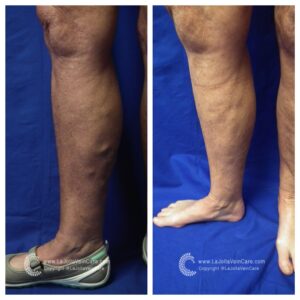
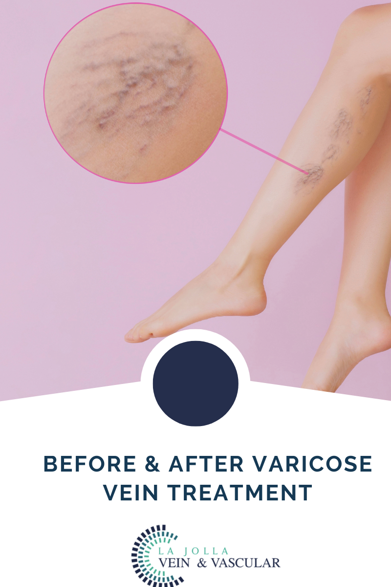
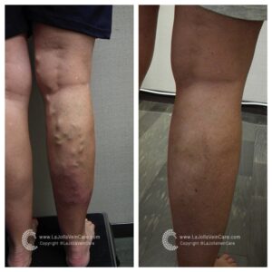
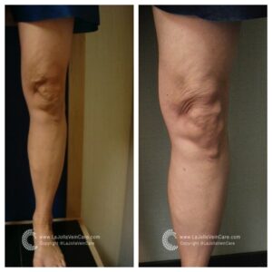
As you can see below, these patient transformations after varicose vein treatment are remarkable!

varicose veinvaricose veinvaricose vein


La Jolla Vein Care Before and After Transformations:


At La Jolla Vein Care, we are here for our patients from beginning to the end. We love seeing the transformations that take place. Below are a few cases from our patient transformations from before varicose vein treatment to after varicose vein treatment. 


If you experience any vein disease symptoms, please call our office at (858)-550-0330 to schedule a consultation with one of our knowledgeable doctors at La Jolla Vein and Vascular. 


For more information on vein health please check out our Youtube Channel or visit our helpful guide of resources. 


Before & After Varicose Vein Treatment2022-12-13T13:08:14-08:00

20, 6, 2022

Pelvic Venous Congestion Treatment

2022-05-23T15:15:40-07:00

Pelvic Venous Congestion (PVCS) Treatment

What are the treatment options for Pelvic Venous Congestion (PVCs)? Available treatment options for PVCs include pain-relieving progestin hormone drugs, ovarian function-blocking Gonadotropin-releasing hormones, vein embolization, and surgery to rid of damaged veins, uterus, and ovaries. Customarily, your doctor will start you on the medications first to relieve pain. If the medications fail, then they may advise on the next step, usually the invasive options. Vein embolization is the most commonly used procedure. Here’s a look at what it involves:

-Pelvic Venous Embolization

-Sclerotherapy

-Radiofrequency Ablation (RFA)

pelvic venous congestion


Pelvic Venous Embolization

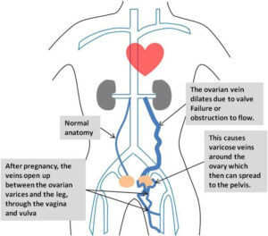
Treating PVCS diseases like pelvic and labial varicose veins requires resolving the backward flow of blood in the ovarian and pelvic veins through embolization. Embolization is an outpatient procedure that uses catheter technology to access the venous system to the problematic ovarian veins. The procedure is painless and requires no major incisions. The physician embolizes each problem vein (blocking blood flow) with vascular plugs or coils. Doing so permanently seals off the varicose veins, diverting blood flow up through healthy veins. The physician will also deliver sclerosing medication to lower the pressure in the pelvic veins and relieve the pain. Once blood can flow smoothly through healthy veins in the pelvic and labial area, the pain disappears. Patients can usually return to work and light activities the day following the procedure and see a full recovery in about one week; at least 7 in 10 women who undergo the procedure report feeling better. This procedure is usually performed by a trained interventional radiologist in the X-ray department.


Sclerotherapy

Sclerotherapy is a minimally invasive procedure that injects sclerosing solutions into the veins. It is used to treat spider, reticular and varicose veins. It also blocks the unsightly or non-functioning veins to gradually improve appearance. This generally involves multiple treatments over the course of several months for pelvic venous congestion. 

Radiofrequency Ablation is a minimally invasive, image-guided procedure used to treat varicose veins. An RFA utilizes high-frequency radio waves directed through a thin tube to create intense heat within the varicose vein. This closes up the problem vessel so blood cannot flow through it, redirecting blood flow into healthy veins to allow the blood to return to the heart. Eliminating the unhealthy vein reduces the vein bulging and minimizes leg pain and swelling. It is performed as an outpatient procedure with minimal recovery and discomfort.


When Should I Contact My Doctor?

Some women have enlarged veins with no symptoms; others have enlarged veins that cause pains and aches. In most cases, these don’t require a medical emergency. The symptoms should reduce as you head into menopause. However, in case of chronic, unbearable pain, nothing is stopping you from seeking medical help. In fact, see your healthcare giver right away for help.

To get the most out of the pelvic venous congestion consultation, here’s what to do:

-Know the purpose of the visit and what you want to get from it

-Before visiting the doctor, put down questions you need them to answer.

-Bring company along to help ask questions and note down points from the doctor.

-Write down the name of the diagnosis, its tests, and treatment options explained by the doctor. -Also, take notes of any new instructions during each visit.

-Know the reasons for new medication prescriptions, their benefits, and their risks.

-Inquire whether it’s possible to treat your condition another way

-Know why and what the results of a test procedure mean

-Ask what will happen if you don’t undergo testing or take medication.

-Ask whether a follow-up appointment is available and note down the visit’s time, date, and reason.

-Ask the doctor for their contacts in case you need a further chat.


 For more information on pelvic venous congestion and to book a consult, please give our office a call at 858-550-0330.

For more information please check out our Youtube Channel at this link.

Pelvic Venous Congestion Treatment2022-05-23T15:15:40-07:00

Arterial Treatments

2022-05-23T14:56:03-07:00

Arterial Treatments


Peripheral Arterial Disease – Treatment

Arterial treatments for the peripheral arterial disease (PAD) has two main goals. One is to manage the symptoms, including leg pain, allowing you to resume your normal physical activities. The second one is to top the progression of atherosclerosis in the body, which reduces the risk of stroke and heart attack.

The good news is that through early intervention, you can achieve these goals through lifestyle changes. Quitting smoking is one of the most significant things you can do to stop the progression of Pad. Walking and exercising regularly following supervised exercise training can improve your symptoms. However, if your peripheral artery disease is at an advanced stage, you will need additional medical treatment. Here are some of the standard peripheral artery disease treatment options available.

Atherectomy

A lower extremity atherectomy is a minimally invasive surgical procedure involving a catheter to remove plaque from arteries. The doctor usually makes a small incision in the artery to insert the catheter. The procedure is performed under local anesthesia to prevent pain. A catheter collects removed plaque in a chamber placed at its tip, ensuring that all the particles are removed from the artery. Depending on the amount of plaque in your arteries, your doctor will repeat the procedure several times to ensure optimal blood flow.

Why Do I Need This Procedure?

The primary goal for the atherectomy is to eliminate plaque buildup in the artery. You may need this procedure when your arteries become too narrowed or blocked due to increased plaque. When the arteries are blocked, blood flow is restricted, meaning that your tissues will not have sufficient oxygen and nourishment they need to survive. Insufficient blood flow causes the muscle in your feet to cramp and lose strength. The procedure is not ideal for all people. Before performing the procedure, the doctors customize to meet the patient’s unique needs. 

Where is the Procedure Performed?

We usually perform the procedure in our facility. Our board of certified doctors have undergone rigorous training to ensure the best possible results. We also have a state-of-the-art facility with modern atherectomy tools that ensure success even for complex conditions. Our doctors also have a vast wealth of experience in this field. Having performed thousands of atherectomy procedures is sure that you are in the right hands. 

How do I Prepare for an Atherectomy?

A few days before the procedure, the doctor usually performs a pre-procedure test to ensure that it is safe for you to undergo the procedure. The doctor may recommend discontinuation of using certain medications days before the procedure. Our healthcare team will also provide you with specific instructions to prepare for the procedure.


Arterial Treatments2022-05-23T14:56:03-07:00

31, 5, 2022

What is Uterine Fibroid Embolization (UFE)

2022-04-29T17:29:18-07:00

What is Uterine Fibroid Embolization (UFE)

With modern medication and care, conditions like fibroid tumors have become more treatable and easier to manage. Uterine Fibroid Embolization (UFE), for instance, is a fairly straightforward procedure used in the treatment of fibroid tumors in the uterus.

Sometimes referred to as uterine artery embolization (UAE), the uterine fibroid embolization (UFE) procedure helps countless women plagued by fibroid tumors.

What does the uterine fibroid embolization treatment entail, and how can you prepare for the procedure? Here’s everything you need to know about uterine fibroids treatment.

What is Uterine Fibroid Embolization?

The muscular walls of the uterus can sometimes be invaded by non-cancerous fibroid tumors. These tumors are also known as myomas and are a form of benign tumors. While these fibroid tumors rarely become cancerous, they can lead to other severe conditions such as increased menstrual bleeding, bowel or bladder pressure, and pelvic discomfort. 

Admittedly, while uterine fibroid embolization shows a high success rate, more research is still needed on the subject. For instance, at the moment, the procedure is usually performed on women who no longer desire to get pregnant. The UFE procedure may also be used on women who wish to avoid a hysterectomy procedure. 

When is Uterine Fibroid Embolization Used? 


As mentioned above, uterine fibroid treatment is rather straightforward and minimally invasive. As a result, the procedure can be used in a wide variety of cases. Some of the instances where the treatment is necessary or advocated for include;

  • Malignant gynecological tumors
  • Hemorrhage after childbirth
  • General or specific trauma


How to Prepare for a Uterine Fibroid Embolization Procedure


The healthcare professional will first need to ascertain if the fibroid tumors are the underlying cause of any visible and diagnosable symptoms. Afterward, they’ll need to determine the size, location, and the number of fibroids. This can be done with the help of an ultrasound machine or magnetic resonance imaging (MRI) equipment.

Moreover, your gynecologist may also need to run a physical examination through a laparoscopy examination. This test gives a clearer picture of the uterus and the invasive fibroids. You could also have a biopsy of the inner lining of the uterus if you are bleeding excessively in between your periods. The procedure known as endometrium helps rule out cancer as the cause of bleeding.

If you are on any medication, supplements, or herbal remedies, you’ll need to inform the doctor before the procedure. Additionally, make a list of any known allergies, including general and local anesthesia and the dye or contrast materials.

It’s also wise to notify the healthcare team if you have recently suffered an illness, just finished treatment, or are on other medication. For example, if you’re on blood thinners, the healthcare provider may advise you temporarily halt the medication in preparation for the procedure.

Women who suspect they are pregnant should let the doctor know well before the uterine fibroid embolization treatment. Some imaging tests are usually not performed during pregnancy in a bid to minimize any radiation exposure to the fetus. In the case of an imaging test, such as an x-ray, which is essential to the operation, particular care guidelines must be followed to manage and reduce radiation exposure.

While preparing for the procedure, you’ll likely be put off solid meals after midnight to the procedure’s day. While the procedure takes a few hours, the doctor could request to stay overnight for further observation.


What Equipment to Expect


Since this is a minimally invasive procedure, the tools and equipment used are reasonably common and risk-free. Some of the equipment to be used in uterine fibroid embolization treatment includes a catheter, x-ray machines, and various embolic agents and medications.

The synthetic material used is known as embolic agents and varies in composition and efficacy. The three most common types of embolic agents are;

-Gelfoam whose texture resembles spongy material

-Polyvinyl alcohol, which resembles coarse sand

-Microspheres, which is a polyacrylamide sphere coated with gelatin

These tolls, equipment, and medication are all safe to use during the uterine fibroid embolization treatment.


Benefits and Risks Associated with Uterine Embolization Treatment


Some of the advantages of uterine fibroid embolization include;

-There’s no need for surgery as the procedure is minimally invasive

-You can resume regular activity sooner and faster

-The procedure is highly effective, boasting well over 90% efficacy rating

-Fibroids rarely regrow after the uterine embolization treatment

-On the other hand, some of the risks that could occur during the procedure are;

-The procedure pierces the skin and could leave an infection if not properly cleaned

-The embolic agents need to be guided carefully, or they could lodge in the wrong area

-Fibroids can lead to chronic pain and an array of other conditions. With uterine embolization treatment, you can now get relief and get back to your healthy self.


What To Expect during Uterine Fibroid Embolization Procedure


A UFE usually takes between 1 and 3 hours and patients usually require about 6 hours of bed rest after the procedure. Patients may experience mild discomfort as the embolization takes full effect. The fibroid tumors breaking down and exiting the body may cause light bleeding for the few weeks after the procedure. Most patients can expect to return to normal activity 7 to 10 days after the procedure. Our physicians will most likely recommend a follow-up consultation after the UFE 1 to 3 weeks afterward and an ultrasound or MRI 3 to 6 months later to monitor your progress.
 

What is Uterine Fibroid Embolization (UFE)2022-04-29T17:29:18-07:00

What are the risk factors & treatments for uterine fibroids?

2022-04-29T17:24:18-07:00

Who is at Risk of Developing Uterine Fibroids?


There are certain natural circumstances, habits, and lifestyle choices that have been shown to come with an increased chance of developing uterine fibroids, as follow:

Ethnic Origin

Research indicates that women of African American ethnicity are at a significantly higher risk of developing uterine fibroids than Caucasian women for comparison. 

Age

A woman’s advancing age places her at increased risk of developing uterine fibroids. The condition will mostly be found in women in their mid-late forties or early fifties, where the majority enter or pass through their menopausal years. 

Family History

Simple stated, this means that those with a history of uterine fibroids will be more likely to develop the condition. 

Eating Habits

It is believed that the consumption of red meat (beef, ham, etc) in copious amounts will increase the risks of uterine fibroid development. The consumption of green vegetables, on the other hand, is thought to reduce these odds. 

Obesity

Being overweight is considered to be another variable increasing women’s risk of developing this condition, with overweight patients being two to three times more likely to be affected than the average population. 


Treatment Options for Uterine Fibroids

The varied nature of uterine fibroid cases and how they affect different patients in different ways means that dealing with the condition has numerous different approaches. Consultations between patients, whether presenting with symptoms or not, and their physicians should be able to see them agree on a course of action that will satisfy the patient’s best interests. Aside from medication, the typical treatment for fibroids is traditional surgical procedures. At Pedes Orange County, we offer a minimally invasive treatment option that can be reviewed below:

Traditional Surgical Procedures

These are considered invasive surgeries that involve extensive physical intervention. They include:

  • An abdominal myomectomy is an option mostly employed in cases where very large, very deeply embedded, or multiple fibroids are encountered. An open abdominal procedure is often the second-to-last option for patients who see hysterectomies as a last resort.
  • Hysterectomy: This major surgical intervention involves the entire removal of the patient’s uterus and is the only guarantee against the recurrent development of uterine fibroids afterward. While most women will have the option of retaining their ovaries, some will have to undergo hormone replacement therapy if they are not to enter menopause.

Minimally Invasive Surgical Procedure (Uterine Fibroids Embolization by La Jolla Vein & Vascular)

At La Jolla Vein & Vascular, we specialize in Uterine Fibroids Embolization. This minimally invasive surgical procedure involves destroying fibroid masses without necessarily removing them entirely. The techniques are most commonly applied in clinical settings due to their effectiveness, proven track record, and minimal patient impact.

  • Uterine artery embolization: (Also known as Uterine fibroid embolization). Embolic agents (small particles) will be introduced into arteries to block blood flow through them, thus starving fibroids of the nutrients needed for their survival and growth.

In surgical procedures that do not entirely remove the uterus, there will be a remaining risk of new uterine fibroid development in the future.

Why Uterine Artery Embolization (UFE) Instead of a Hysterectomy

Benefits of UFE Over Hysterectomy

Uterine artery embolization (UFE ) is a minimally invasive procedure that effectively treats uterine fibroids and is considered an alternative to uterine fibroid surgery removal. Uterine artery embolization (UFE) is typically performed as an outpatient procedure, which means that most women go home the same day for recovery.

UFE has several benefits over hysterectomy. Besides involving only a small nick in the groin or wrist for catheter insertion, the uterus is preserved. There is no scar with UFE. For a UFE, conscious sedation is used, unlike a hysterectomy, where the woman receives general anesthesia. The recovery time for UFE is generally within 2 weeks, which is significantly faster than the usual 6-week recovery time after hysterectomy.

Women who have a hysterectomy that removed both the uterus and ovaries usually get estrogen replacement therapy (ERT) alone. But women who have only the ovaries removed need both estrogen and progestin because estrogen alone can increase the risk of cancer in the uterus.

The recovery time for UFE is generally within 2 weeks, which is significantly faster than the usual 6-week recovery time after hysterectomy.

The idea of having your entire uterus removed is psychologically daunting for women as some women may still want the option to carry a child, do not want to undergo a general surgery, and/or feel like removing the uterus takes away part of their identity as a female.

Treatment for Uterine Fibroids

With modern medication and care, conditions like fibroid tumors have become more treatable and easier to manage. Uterine fibroid embolization, for instance, is a fairly straightforward procedure used in…


What are the risk factors & treatments for uterine fibroids?2022-04-29T17:24:18-07:00

What causes uterine fibroids?

2022-04-29T17:20:54-07:00

What Causes Uterine Fibroids?

While the science is not completely settled on what exactly causes or is behind the formation of uterine fibroids in human beings, there are certain factors that researchers believe play significant roles in triggering their growth or development.

Genetics

In 50 percent of cases, patients will be found to have some genetic abnormality. In many instances, some erroneous translocation will be discovered on some chromosomes.

Hormones

While we may not be completely certain as to what lies at the root cause of uterine fibroids, it has been clearly demonstrated that hormones play a large role in determining their growth or progression. While a woman is pregnant, they will see rapid growth due to the presence of the hormones estrogen and progesterone. Should the patient use anti-hormone medication, the opposite will happen – the fibroids will shrink. Furthermore, fibroids will stop growing or begin shrinking once a patient reaches menopause, which is the point at which these hormones also cease being produced in the female body.

uterine fibroids

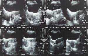
Ultrasound image of lower abdomen, ovary and uterus with tumor or uterine fibroid, leiomyoma of female woman patient for gynecological medical exam, analysis and test

Testing and Diagnosis of Uterine Fibroids

In many cases, especially those whereby uterine fibroids do not present any noticeable symptoms for the patient, this condition will be discovered in a regular pelvic exam. This is a routine checkup carried out by gynecologists. While this checkup might determine the presence of a mass or lump on the uterus, further testing will have to be carried out to make a definite diagnosis.

These testing methods include:

Note: Upon learning that fibroids are a type of tumor, the foremost concern for many patients is whether the tumor is cancerous or might predispose them in some way to developing cancer. Fibroids are rarely cancerous, with the chances of such fibroids (leiomyosarcomas) arising at less than one in a thousand.  Additionally, the presence of fibroids in no way increases a patient’s chances of developing any type of cancer.


What causes uterine fibroids?2022-04-29T17:20:54-07:00

Complications and Causes of Varicoceles

2022-04-29T16:58:30-07:00

What are Varicoceles?

A varicocele is a term used to describe abnormal enlargement of the veins within the scrotum. The veins carry blood from the various body organs back to the heart. Typically, veins have valves that prevent blood backflow. When the valves in the veins within the scrotum don’t work as they should, blood tends to collect in the scrotum, leading to varicoceles.

While varicocele is very common, it is not life-threatening. In fact, research shows that about 15 percent of all adult men have varicocele. For most men, the condition will go unnoticed throughout their life or does not cause any complications. Although the condition is not dangerous, varicocele is a common cause of low sperm production and reduced sperm quality, leading to infertility. Varicoceles can also lead to reduced testosterone production and scrotal discomfort. The condition often does not need treatment until there is a reason for concern about the associated problems. Fortunately, varicocele is easy to diagnose and treat.


Complications and Causes of Varicoceles


Unfortunately, although the condition is not life-threatening, it can also lead to several unpleasant complications.

  • Atrophy (shrinking of the affected testicles)

The testicles are made of sperm-producing tubules. Varicoceles damage these tubules, making the testicles soften and shrink.

  • Infertility

The condition also causes the local temperature around or in the testicles to be too high, affecting sperm formation


Causes

Medical practitioners are not sure of the leading cause of varicoceles. However, doctors agree that the condition forms when there is a problem with blood flow in the spermatic cord. The problem develops when the valves in the veins do not function properly and allows blood to flow back. As a result, blood pools in the veins causing them to dilate. When the condition is common in adults, it can also develop in teens during puberty. The testicles usually require more blood than usual during this growth cycle. This condition can lead to problems in the veins, preventing the blood from flowing where it should.

For more information on vein and vascular treatment please visit our YouTube Channel. 


Complications and Causes of Varicoceles2022-04-29T16:58:30-07:00

Varicocele Diagnosis and Treatments

2022-04-29T16:50:34-07:00

Varicocele Diagnosis


Your doctor will perform a thorough physical examination, which might reveal a non-tender mass above the testicles to diagnose varicoceles. When the mass is large enough, it feels like a bag of worms. When the varicocele is small, the doctor might request you to stand, take a deep breath and hold it while you bear down. This technique will help the doctor to detect any abnormal veins enlargement. Your doctor may also order a scrotal ultrasound. The test uses high-frequency sound waves to create a precise image of the structure of the scrotum. The test is also helpful in helping the doctor rule out other reasons that could be causing the problem in the scrotum.


Treatments for Varicocele

 In most cases, varicoceles treatment is not necessary. Most men with varicoceles do not    experience   fertility issues. However, if the condition is causing pain, testicular atrophy, or infertility, you can benefit.

In most cases, varicoceles treatment is not necessary. Most men with varicoceles do not experience fertility issues. However, if the condition is causing pain, testicular atrophy, or infertility, you can benefit from varicoceles treatment. The treatment helps in sealing off the affected vein and redirecting the blood flow into normal veins. In case of infertility issues, varicoceles treatment helps in improving the quality of the sperm and sperm count. The procedure that we perform at LA Jolla Vein & Vascular on varicocele is percutaneous embolization of varicocele embolization.

The primary goal for treatment procedures is to help prevent the backflow of blood from the body to the scrotum, helping in cooling off the testes. Surgical procedures will help restrict the blood flow to the veins leading from the testis to the body. However, not all the veins are sealed off. The vassal vein, which is not subject to the same problems as the other varicose veins, is left open to allow blood to leave the testicles after the procedure.

Some of the clear indications that you need varicoceles repair during adolescence include pain, progressive testicular atrophy, and abnormal semen analysis results. While this treatment helps in improving sperm characteristics, it is not clear whether untreated varicoceles can lead to progressive sperm worsening. 

The common varicoceles treatment options include (La Jolla Vein & Vascular Only Performs Varicocele Embolization):

Percutaneous Embolization (Varicocele Embolization)

    Varicocelectomy

    Laparoscopic Surgery

    Open Surgery


 Recovery

In most cases, you can go back to your routine after two days of varicose treatment. However, it is advisable to take it easy. Avoid strenuous activities and exercise for about two weeks.People who have undergone percutaneous embolization tend to recover faster. While you may need a day or two off from work, you can return to your workout program in 7-10 days. If the treatment procedure is to help with fertility, your doctor will perform an additional test in 3-4 months. You will be able to see improved results in 6-12 months. It is important to note that more than 50 percent of men who have undergone the procedure have restored their fertility. Additionally, these surgical procedures are also crucial for teens as they help in slowing testicular growth.


Varicocele Diagnosis and Treatments2022-04-29T16:50:34-07:00
Go to Top