La Jolla Vein & Vascular Arterial Exams and Vascular Lab
LJVascular2023-09-29T19:28:20-07:00La Jolla Vein & Vascular Arterial Exams and Vascular Lab
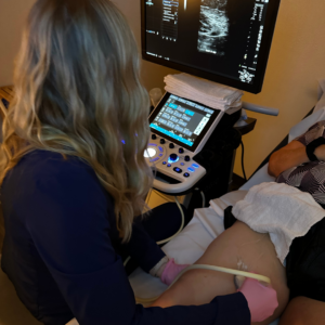
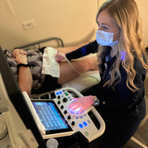
When it comes to vascular health, early detection and accurate diagnosis are paramount. At our clinic, we offer a one-stop solution for vascular imaging and physician consultations, all under one roof. With a state-of-the-art non-invasive vascular laboratory, we utilize advanced imaging technology to detect and assess vascular diseases that impact blood flow in the arteries and veins. In this blog post, we’ll explore the convenience and expertise of our vascular lab, the conditions we diagnose, and the types of ultrasound testing we provide.
The Advantages of Our Vascular Laboratory
Our commitment to your vascular health extends beyond traditional healthcare. We provide a range of benefits to ensure your comfort and convenience:
- Non-Invasive Testing: Our vascular laboratory specializes in non-invasive testing methods, ensuring a comfortable experience for our patients.
- Same-Day Imaging Appointments: We understand the urgency of timely diagnosis. That’s why we offer same-day imaging appointments to promptly address your vascular concerns.
- Comprehensive Care in One Location: Say goodbye to the hassle of visiting multiple locations for imaging, diagnosis, and treatment. At our clinic, you can undergo vascular imaging and consult with a healthcare provider all in the same place.
- Comfortable, Private Rooms: Your comfort is our priority. Our private examination rooms are designed to provide a tranquil environment for your vascular tests.
Conditions We Diagnose
Our vascular laboratory is equipped to diagnose a wide range of vascular conditions, including but not limited to:
Vein Diseases:
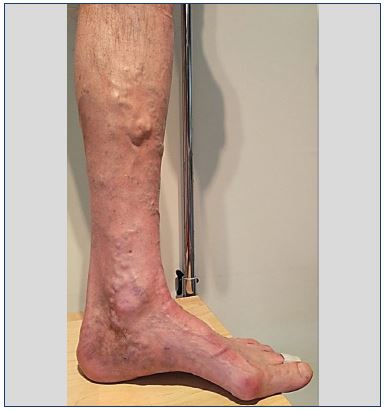
- Varicose Veins
- Chronic Venous Insufficiency
- Deep Vein Thrombosis (DVT)
- Carotid Artery Disease and Stroke (TIA or Stroke)*
Artery Diseases:
- Peripheral Arterial Disease (PAD)**
- Abdominal Aortic Aneurysm (AAA)
- Upper Extremity Arterial Disease
Understanding Duplex Ultrasound
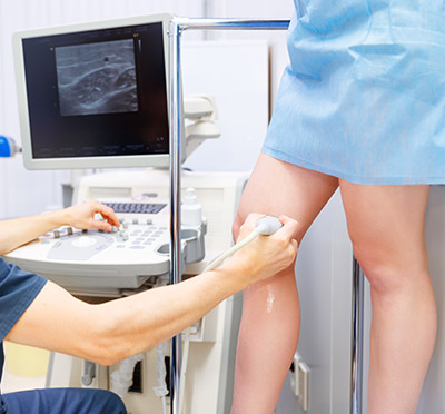
Duplex ultrasound is a powerful diagnostic tool used in our vascular laboratory. It combines Doppler flow information with conventional imaging (B-mode) to provide a comprehensive view of your blood vessels. Key aspects of duplex ultrasound include:
- Structure Visualization: Duplex ultrasound allows us to see the structure of your blood vessels, including the diameter and any obstructions.
- Blood Flow Assessment: It determines how fast blood moves through your vessels, assessing the direction of blood flow and identifying any blockages or blood clots.
- Valve Function: The technology also examines the function of valves within your veins and arteries.
- Deep Vein Assessment: Duplex ultrasound can visualize deep veins within the muscles, providing crucial information for diagnosing conditions like varicose veins.
Types of Ultrasound Testing Offered
Our vascular laboratory offers a variety of ultrasound testing options, including:
Direct Testing (Duplex Imaging):
- Venous Exams for Deep Vein Thrombosis and Venous Reflux
- Arterial Exams for Abdominal Aorta, Abdominal Aortic Aneurysm (AAA) Screening, and Carotid Duplex
- Lower Extremity Duplex
Indirect Testing (Non-Imaging):
- Arterial Testing such as Ankle Brachial Index (ABI) with waveforms and Segmental pressures with waveforms (P&Ws) for upper or lower extremity
Venous Reflux or Venous Insufficiency Assessment
Our Duplex Ultrasound examination allows us to visualize blood vessels that are not visible to the naked eye, even deep within the muscles. This examination is crucial for assessing the underlying condition causing varicose veins. By identifying veins with faulty valves and mapping their anatomy, we create a roadmap for an accurate assessment and an effective treatment plan.
Before Your Test
Preparing for our ultrasound study is simple. Just remember these key points:
- No preparation is required for this study.
- Avoid wearing compression stockings on the same day as the examination.
- Stay hydrated for the best experience.
Our vascular laboratory is dedicated to providing you with top-notch vascular imaging services in a convenient and comfortable environment. By offering non-invasive testing, same-day appointments, and a comprehensive approach to vascular health, we aim to ensure your well-being and peace of mind. Trust us with your vascular concerns, and let us help you on your journey to better health.
“Bringing Experts Together for Unparalleled Vein and Vascular Care”
La Jolla Vein & Vascular (formerly La Jolla Vein Care) is committed to bringing experts together for unparalleled vein and vascular care.
Nisha Bunke, MD, Sarah Lucas, MD, and Amanda Steinberger, MD are specialists who combine their experience and expertise to offer world-class vascular care.
Our accredited center is also a nationally known teaching site and center of excellence.
For more information on treatments and to book a consultation, please give our office a call at 858-550-0330.
For a deeper dive into vein and vascular care, please check out our Youtube Channel at this link, and our website https://ljvascular.com
For more information on varicose veins and eliminating underlying venous insufficiency,
Please follow our social media Instagram Profile and Tik Tok Profile for more fun videos and educational information.
For more blogs and educational content, please check out our clinic’s blog posts!


