14, 12, 2023

Venous Insufficiency: When Your Veins Need a Helping Hand

2023-10-11T16:30:21-07:00

Venous Insufficiency: When Your Veins Need a Helping Hand

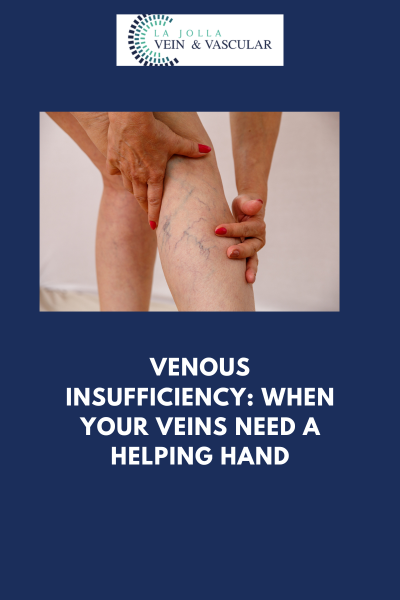
Venous reflux disease, sometimes referred to as venous stasis, venous insufficiency, or venous incompetence, stands as a multifaceted condition that intricately affects the veins in the legs. In this article, we delve into the intricacies of venous reflux disease, elucidating its causes and symptoms, and shedding light on the progressive nature of this ailment. We’ll also delve into the indispensable role played by ultrasound technology in the diagnostic process and the formulation of tailor-made treatment plans.

Unraveling Venous Reflux

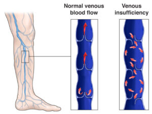
At the core of venous reflux disease lies the concept of ‘leaky valves’ within the leg veins. These valves, entrusted with the responsibility of maintaining the smooth flow of blood, may falter, permitting blood to flow backward (a condition known as reflux) rather than progressing toward the heart. Venous reflux has the potential to manifest in both deep and superficial leg veins, subsequently influencing the efficiency of blood circulation.

Demystifying the Anatomy of Reflux

Within the realm of leg veins, there exist two primary categories: deep and superficial. The deep veins, nestling within the muscular tissue, shoulder the responsibility of transporting the lion’s share of blood from the legs back to the heart. Conversely, superficial veins are situated just beneath the skin, external to the muscle. Among the pivotal players in the realm of superficial veins are the Great Saphenous Vein (GSV), meandering through the thigh and calf, and the small saphenous vein, traversing the back of the calf.

The Ramifications of Leaky Valves

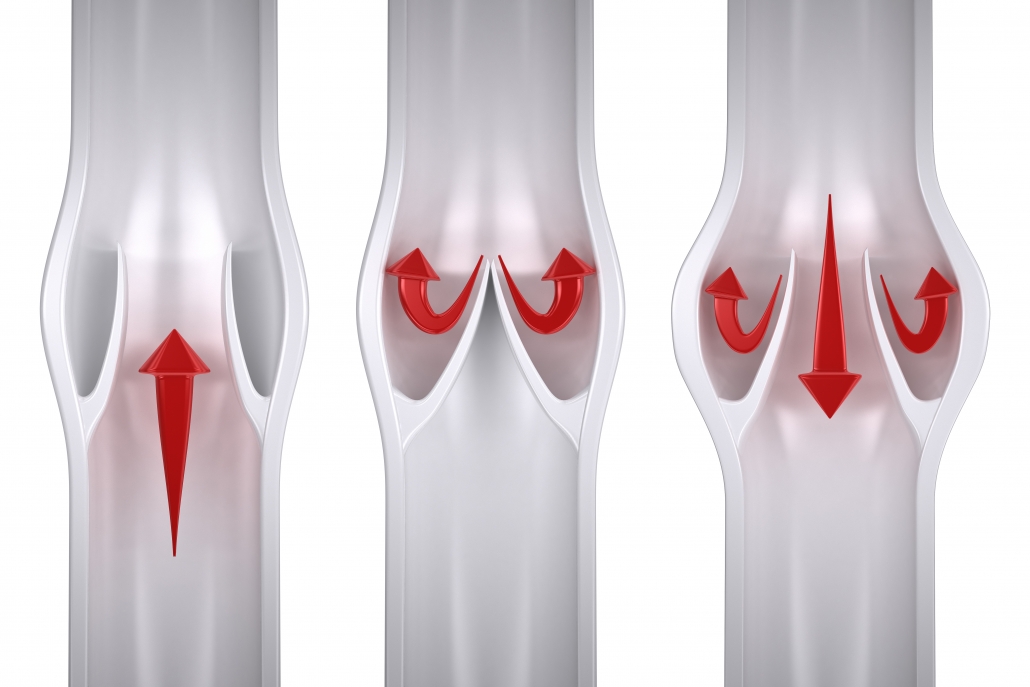
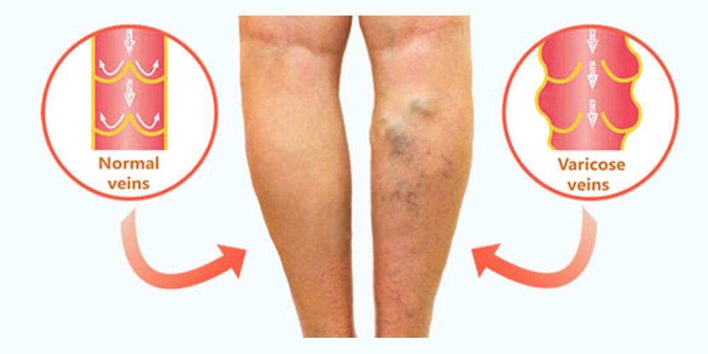
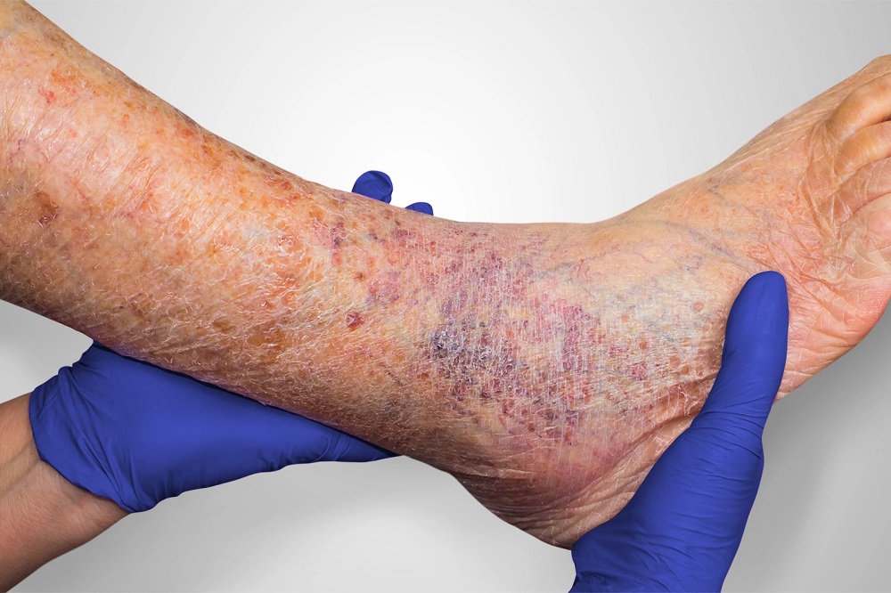
Under normal circumstances, one-way valves present in leg veins facilitate the upward flow of blood against the force of gravity, aided by the rhythmic contractions of calf muscles. However, when these valves succumb to leakage, the flow of blood reverses, leading to the accumulation of blood in the lower legs. This condition is accompanied by a medley of distressing symptoms, including the sensation of heavy legs, pain, fatigue, ankle swelling, and even restlessness in the legs during the night. As time unfolds, venous reflux disease can advance, resulting in skin changes such as darkening, dryness, itching, and the potential emergence of venous leg ulcers.

The Role of Ultrasound in Diagnosis

The diagnosis of venous reflux disease necessitates specialized tools, with ultrasound technology taking center stage. Not all vein-related issues are discernible to the naked eye, as many originate from veins concealed beneath the skin’s surface. Ultrasound examinations grant healthcare professionals valuable insights into the direction of blood flow, the functionality of valves, and the presence of any obstructions or scars within the veins.

A Tailored Approach to Treatment

Effectively addressing venous reflux disease requires a strategic and customized approach, recognizing the unique characteristics of each patient’s condition. The treatment process generally encompasses three fundamental steps:

Step 1: Rectifying Underlying Reflux

The initial focal point revolves around tackling the root cause—venous reflux. This is achieved by targeting the saphenous veins, which often serve as the epicenter of the issue. Cutting-edge vein ablation procedures, including radiofrequency ablation, laser ablation, mechanico-chemical ablation (MOCA), and Varithena Foam, are deployed to restore the standard flow of blood.

Step 2: Combating Varicose Veins

Once the underlying reflux has been successfully addressed, the focus shifts to addressing varicose veins. Techniques like foam sclerotherapy, involving the injection of a foamed medication, or minimally invasive removal methods, are utilized to eliminate bulging veins.

Step 3: Managing Spider Veins

For those desiring cosmetic enhancements, the treatment of spider veins through sclerotherapy is available. Although primarily cosmetic, this step serves to conclude the all-encompassing treatment journey.

Venous reflux disease presents as a multifaceted condition that demands specialized care for effective management. Our comprehensive approach encompasses state-of-the-art diagnostics, cutting-edge treatments, and patient-tailored care to address the various facets of this ailment. Through our expertise and unwavering dedication, our aim is to deliver transformative outcomes, ultimately enhancing the health and quality of life of our patients. If you are prepared to embark on the path to healthier veins, do not hesitate to reach out to us to take the first stride toward comprehensive vein and vascular wellness.

“Bringing Experts Together for Unparalleled Vein and Vascular Care”

La Jolla Vein & Vascular (formerly La Jolla Vein Care) is committed to bringing experts together for unparalleled vein and vascular care. 

Nisha Bunke, MD, Sarah Lucas, MD, and Amanda Steinberger, MD are specialists who combine their experience and expertise to offer world-class vascular care. 

Our accredited center is also a nationally known teaching site and center of excellence. 

For more information on treatments and to book a consultation, please give our office a call at 858-550-0330. 

 For a deeper dive into vein and vascular care, please check out our Youtube Channel at this link, and our website https://ljvascular.com

For more information on varicose veins and eliminating underlying venous insufficiency,

Please follow our social media Instagram Profile and Tik Tok Profile for more fun videos and educational information. 

For more blogs and educational content, please check out our clinic’s blog posts

Venous Insufficiency: When Your Veins Need a Helping Hand2023-10-11T16:30:21-07:00

2, 6, 2023

Patient Transformation: Spider Veins

2023-02-15T20:42:37-08:00

What are spider veins?

Spider veins are the fine, thread-like reddish veins at the surface of the skin. These veins are not healthy. Veins are often considered a cosmetic issue, but they can be associated with underlying feeder veins, not visible to the naked eye.  Feeder veins are the ‘blue veins’ also known as ‘reticular veins,’ that give rise to spider veins. Spider veins may also be a sign of underlying venous insufficiency. For example, spider veins located in the inner ankles and inner thighs can indicate an underlying problem with the saphenous vein. As a result, an ultrasound examination may be recommended to identify and effectively treat the underlying source of these veins.

What are the symptoms?

Vein conditions affect people differently. For some, these veins can be a painless cosmetic concern, and for others they may cause symptoms. The most common symptoms of spider veins are burning, throbbing, and localized pain. They can also feel hot and itchy and bleed.

What causes them?

Spider veins in the legs are caused by the same condition that causes varicose veins. Leaky vein valves allow blood to pool within the veins causing them to stretch and become enlarged. These veins on other areas of the body, such as the face, and chest can be caused by sun damage, hormone changes or liver disease. Hormones, such as with pregnancy, birth control or hormone replacement therapy can weaken the vein wall.

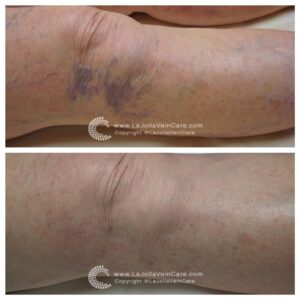
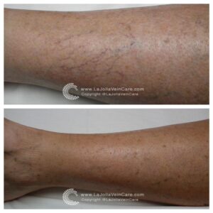
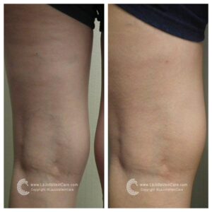
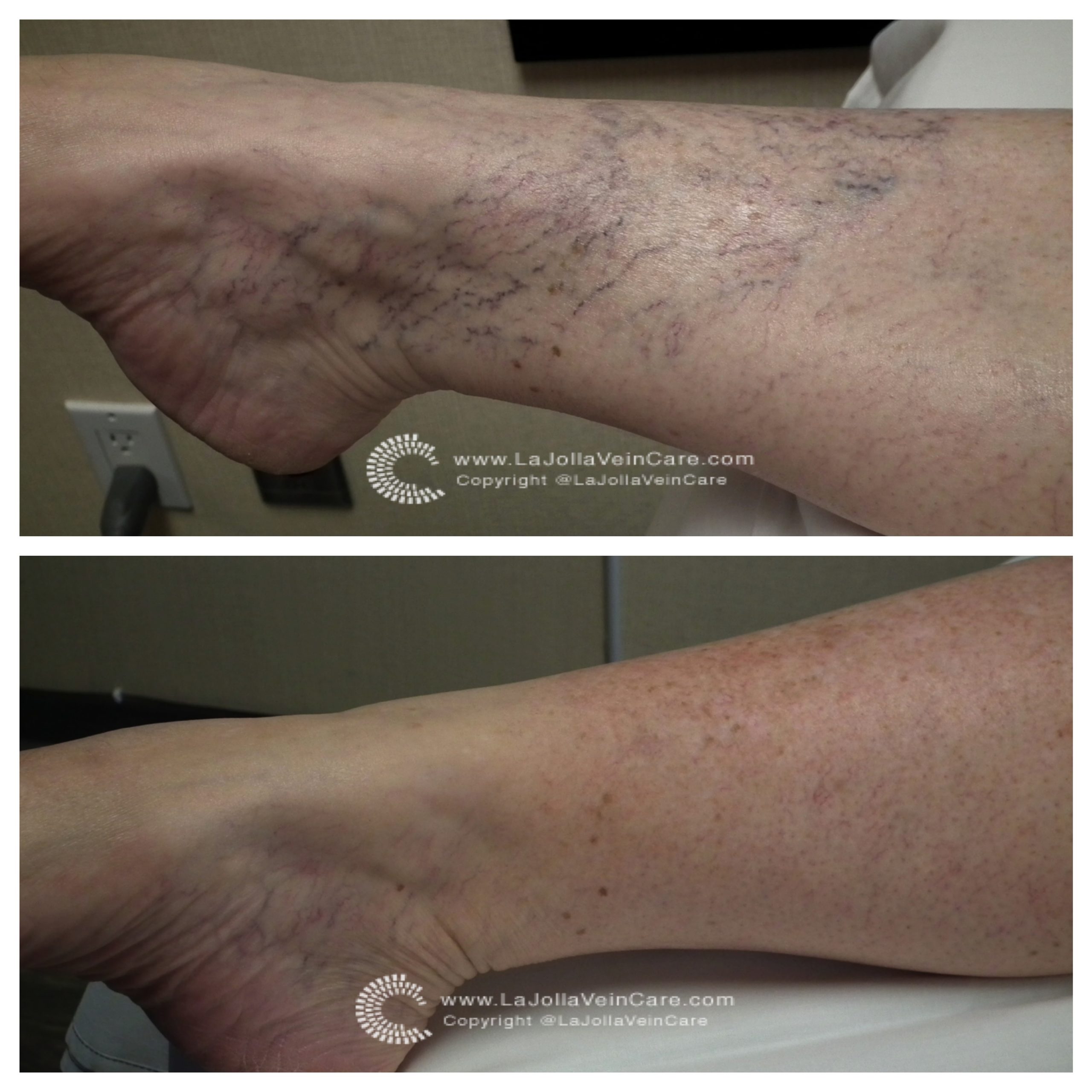
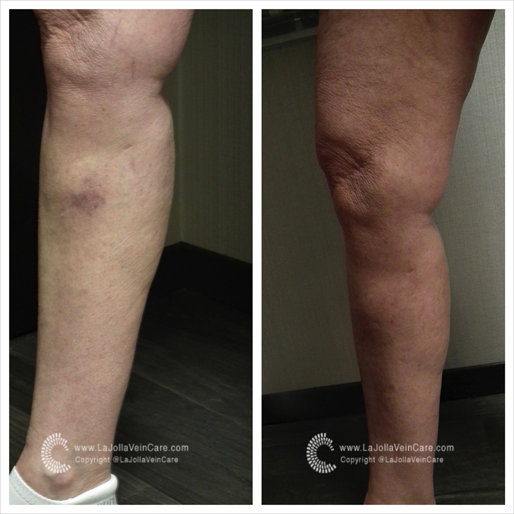
Below you can see the transformation of 3 of our patients who received vein treatment.

spider veins spider veins 7 BA SV spider 13after fotor 1

“Bringing Experts Together for Unparalleled Vein and Vascular Care”

La Jolla Vein & Vascular (formerly La Jolla Vein Care) is committed to bringing experts together for unparalleled vein and vascular care. 

Nisha Bunke, MD, Sarah Lucas, MD, and Elliot DeYoung, MD are specialists who combine their experience and expertise to offer world-class vascular care. 

Our accredited center is also a nationally known teaching site and center of excellence. 

For more information on treatments and to book a consultation, please give our office a call at 858-550-0330. 


For a deeper dive into vein and vascular care, please check out our Youtube Channel at this link.


For more information on varicose veins and eliminating underlying venous insufficiency, check this link out full of resources. 

Please follow our social media Instagram Profile and Tik Tok Profile for more fun videos and educational information. 


Patient Transformation: Spider Veins2023-02-15T20:42:37-08:00

21, 4, 2022

Understanding Venous Stasis Dermatitis

2022-04-15T15:36:58-07:00

Understanding Venous Stasis Dermatitis

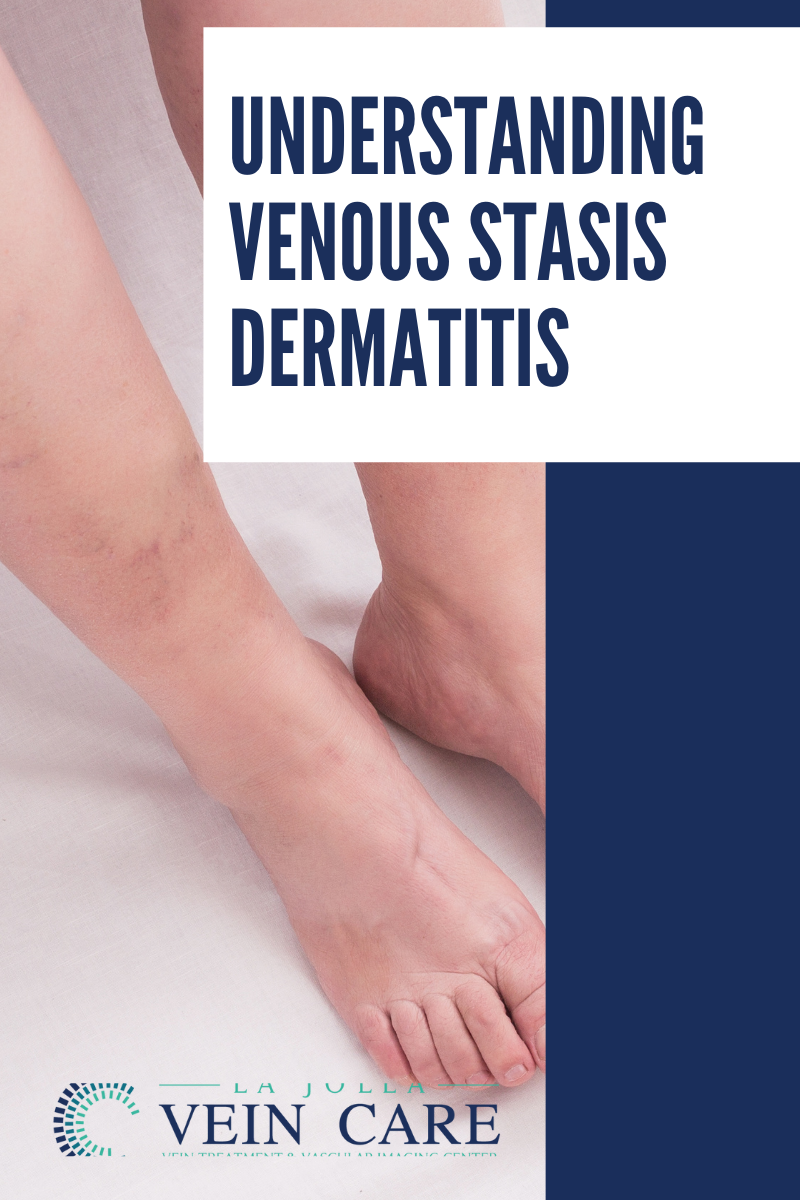
Venous stasis dermatitis is a change in the skin that occur when blood collects (pools) in the veins of the lower leg. ‘Stasis’ refers to pooling of the blood in the lower legs from venous insufficiency, and ‘dermatitis’ refers to the inflammation and related skin changes. Because of the inflammation, the skin around the ankles is usually itchy and discolored.

In venous stasis dermatitis, the skin of the ankles and lower legs may look thin or tissue-like. You may slowly get brown stains on the skin.The skin may become irritated or crack if you scratch it. It may also become red or swollen, crusted, or weepy.Over time, some skin changes become permanent. Thickening and hardening of the skin on the legs and ankles (lipodermatosclerosis)

Here’s what to look out for:

  • A bumpy or cobblestone appearance of the skin
  • Dark brown color

Venous stasis can cause skin sores (ulcers) may develop (called a venous ulcer or stasis ulcer). These most often form on the inside of the ankle. For this reason, skin changes are noticed around the ankle, you should see your doctor or vein specialist to stop the progression of venous insufficiency and prevent the skin from forming an ulceration.

  Stasis dermatitis

Chronic venous insufficiency over time can lead to skin changes and eventually ulceration. This image demonstrates stasis dermatitis (also known as venous dermatitis). Stasis dermatitis refers to the skin changes including skin discoloration around the ankles, dry itchy skin that can be thin, and it may eventually break down to cause an ulceration. You need to see a doctor if these signs are present.

Eight symptoms to watch out for:

  1. Swelling around ankles
  2. Heavy legs
  3. Varicose Veins, which can be twisted, bulging, dark, or purple
  4. Itching
  5. Pain
  6. Sores that ooze, crust or become scaly
  7. Thickened skin around ankles or your shins
  8. Hair loss on ankles and shins
Understanding Venous Stasis Dermatitis2022-04-15T15:36:58-07:00

10, 3, 2022

Understanding Venous Insufficiency

2022-12-13T14:11:01-08:00

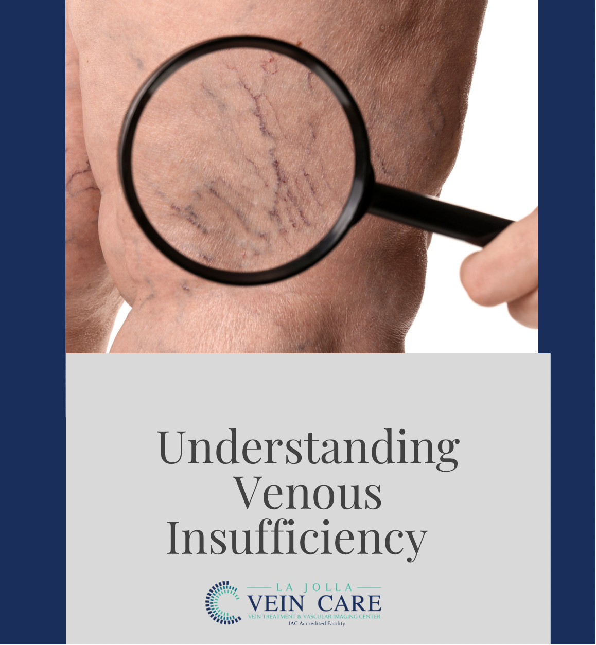
When you become a patient with La Jolla Vein Care, you will go through a screening process, and a vascular evaluation using ultrasound technology to diagnose your venous reflux disease. Chronic venous insufficiency is a disease affecting at least 50 million Americans. It is very common, but unfortunately undiagnosed. Through our venous insufficiency Duplex Ultrasound test, our vein specialists can determine exactly what is going on in the patient’s veins. 

The anatomy and process of venous insufficiency or venous reflux disease: 

One of the major veins that takes the blood to the heart is deep inside the muscles of the thigh and calf called the deep veins. Parallel to this lies the superficial veins inside the legs. Superficial veins dump the blood into the deep vein in the groin area and collectively they take the blood back to the heart. 

The direction of the blood flow begins from the ground up towards the heart. Both deep and superficial have a structure system known as the valves. They function as a safety unit directional valve system. Where the blood can flow upward and not downward and be open or closed off. Similarly the same type of structure is found in your heart with heart valves. 

Over the course of the years, depending on your occupation, family history, pregnancy, surgeries, any trauma or injury to your legs, and any kind of underlying muscular or skeletal problem will contribute to problems with the valves in your legs. The valves, over the course of the years may get farther apart from one another. They will still be able to open and close, but the main problem will be the blood flow will not be able to go in one direction. This causes the veins to become enlarged. As a result,  the valves remain open and the blood to flow upward and also downward. This is known as venous reflux disease or venous insufficiency. 

venous reflux disease

A consultation with one of our vein specialists for chronic venous insufficiency involves taking a detailed history and physical examination in combination with a venous reflux ultrasound study.  Based on your personal findings, a customized treatment plan will be discussed which may include doing nothing, conservative management, or various treatment options based on your individual needs. 

For any more questions about venous disease and the process of diagnosis and treatment, please call us at (858)-550-0330.

Understanding Venous Insufficiency2022-12-13T14:11:01-08:00

21, 5, 2020

Leaky Vein Valve Disease and Varicose Veins

2021-11-05T10:56:55-07:00

Venous reflux disease refers to ‘leaky valves in the veins of the legs. Reflux may occur in the deep and/or superficial leg veins. The deep veins are those within the muscle; they bring at least 80-90% of the blood from the legs back to the heart. The superficial veins are outside of the muscle and under the skin. The main superficial veins are the Great Saphenous Vein that courses up the middle of the thigh and calf and the small saphenous vein, which courses up the back of the calf. Normally, there are one-way valves within the leg veins, which help blood flow in one direction: toward the heart. This means blood is traveling against gravity. The calf muscle also helps move blood toward the heart. When vein valves are leaky, blood flows backward (reflux) towards the feet. Blood pools in the lower legs, causing bulging veins at the surface. Symptoms include leg heaviness, leg fatigue, leg pain, ankle swelling, phlebitis (inflamed and painful veins) restless legs at night, and night cramps. Venous reflux disease is progressive and worsens over time. Skin changes may also develop, including darkening of the skin around the ankles. The darkening of the skin is sometimes referred to as venous stasis skin changes. The skin can become dry and itchy (venous eczema). Eventually, the skin can break down causing a wound, called a venous leg ulcer.

Leaky Vein Valve Disease and Varicose Veins

Leaky Vein Valve Disease and Varicose Veins2021-11-05T10:56:55-07:00

20, 11, 2014

What Are Vein Valves?

2021-11-04T14:10:40-07:00
GSV valve 2

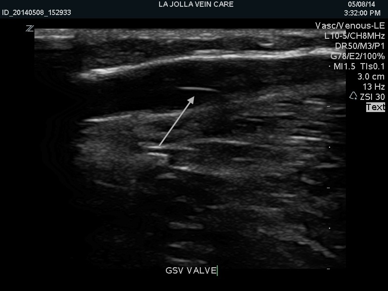
The valves inside the leg veins can be seen on ultrasound. The arrow points to a valve in the great saphenous vein within the leg.

healthy_and_diseased_valves

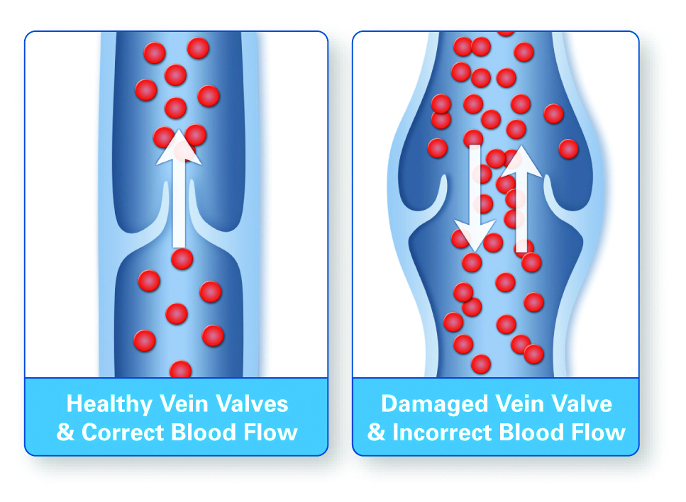
Leg veins have one-way valves that prevent blood from flowing backward. Diseased valves are ‘leaky’ and allow blood flow both forward and backward, eventually causing bulging of the veins, i.e., varicose veins.

In the circulatory system, the veins carry de-oxygenated blood back to the heart. The leg veins carry blood toward the heart, against gravity. Therefore, the leg veins have one-way valves the prevent backflow of blood. When the valves do not function properly, they allow blood to flow backward, causing the pooling of blood. This is referred to as venous reflux or venous insufficiency. Eventually, the backflow of the blood will cause varicose veins to develop and symptoms related to the increased pressure in the leg veins such as leg heaviness, aching, swelling, restless legs, night cramps, throbbing, and pain. 

What Are Vein Valves?2021-11-04T14:10:40-07:00

26, 6, 2014

8 Warning Signs of Vein Disease: #2 Discoloration of the Skin

2021-11-03T17:41:45-07:00

Discoloration of the Skin

Over time, leakage of the blood into the area surrounding the veins can cause tissue to die. The resulting pooling of blood in the tissue causes a darkening of the skin. It is at this stage that the skin is actually stained by your own blood.  This is also known as venous stasis skin changes, skin hyperpigmentation, or venous stasis dermatitis because the skin is often inflamed, itchy, and eczema-like.  This is most frequently caused by venous reflux, an underlying condition that can be treated. When skin changes like these are present, the condition is termed chronic venous insufficiency. Over time, the skin condition may worsen and the skin may become darker, firm to touch, scaly, and itchy, and the skin may break down causing a venous leg ulcer.

8 Warning Signs of Vein Disease: #2 Discoloration of the Skin2021-11-03T17:41:45-07:00

What are Vein Valves?

2021-11-04T19:47:30-07:00
healthy_and_diseased_valves

In healthy veins, the valves close after the blood flows towards the heart, preventing backflow.
When veins become dilated, the valves cannot close properly, allowing blood to flow back towards the ankle.

Vein valves play a critical role in helping blood flow through the veins back to the heart. Like swinging doors, valves open to allow blood to flow toward the heart and flap closed again to prevent the flow of blood back down the legs. If the veins become dilated, the flap-like valves cannot completely close, making them incapable of preventing the backflow of blood. This ‘back flow’ of blood through dysfunctional valves is called venous reflux or venous insufficiency.

What are Vein Valves?2021-11-04T19:47:30-07:00

Why Didn’t My Spider Veins Go Away?

2021-11-04T19:29:17-07:00
1

Before and After spider vein treatment at La Jolla Vein Care. This picture demonstrates that spider veins (reddish spider-like vessels) can stem from underlying feeder vessels (the blue vein). The feeder vein can be compared to the ‘root’ of a weed; If you clip the surface branches of a weed without pulling the root of the problem, the weed will never go away.

DSCF3580_fotor

Veins in certain locations, like in the inner knee or inner thighs and inner ankles, often arise from underlying venous incompetence of a vein called the great saphenous vein that runs from the groin along the inner thigh to the ankle. If spider veins do not respond to treatment, it is important to have an ultrasound evaluation to determine if there is an underlying source of the problem.  Once the underlying problem is properly identified, it can be treated.

Spider veins can be the ‘tip of the iceberg’ and represent a bigger problem, that is not visible to the naked eye. For example, as seen in the first image, spider veins can arise from blue ‘feeder’ veins, which must be treated for best results.

Spider veins can also stem from an underlying problem such as venous reflux in larger veins, such as the great saphenous vein. As demonstrated in the second picture, spider veins in the inner knee, inner ankle and inner thighs can originate from this vein. This can only be determined by ultrasound examination. It is important to have your vein evaluated by a trained specialist. Effective treatment depends on an accurate and thorough diagnosis.

Why Didn’t My Spider Veins Go Away?2021-11-04T19:29:17-07:00

25, 3, 2014

What is a Venous Insufficiency / Duplex Ultrasound Test?

2021-11-04T00:54:25-07:00

The Duplex Ultrasound examination, also called a venous insufficiency study allows us to visualize the blood vessels that are not visible to the naked eye, even blood vessels that are deep within the muscles.  The ultrasound examination is used to both identify the veins that have faulty valves and to map the anatomy of the veins, creating a ‘road map.’  This is necessary to make an accurate assessment of the cause and extent of the varicose veins, as well as to formulate the best treatment plan.  This should be done for any individual being evaluated for varicose veins, leg swelling, patients who have failed prior treatment, patients who are symptomatic and in some patients with certain anatomic patterns of spider veins.

Duplex ultrasound uses sound waves to get images of your blood vessels.  It also helps determine how fast blood moves through the vessels.

Darkening of skin, and itchiness are a sign of chronic venous insufficiency or venous reflux disease

Darkening of skin, and itchiness are a sign of chronic venous insufficiency or venous reflux disease

At La Jolla Vein Care, the venous duplex study will be performed by one of our registered vascular technologists, who have specialized training in the science of vascular sonography (RVT). It is performed in the standing position, which is important in obtaining an accurate study result.  This examination takes approximately 45-60 minutes. Following the study, the doctor will personally discuss the results with our patients to ensure a complete understanding of the ultrasound findings.  Insurance typically covers this type of diagnostic study.


What is a Venous Insufficiency / Duplex Ultrasound Test?2021-11-04T00:54:25-07:00
Go to Top