Ultrasound can detect abnormal blood flow and other vascular conditions not visible to the naked eye
We Provide One Stop Vascular Imaging & Physician Consultations in the same Clinic
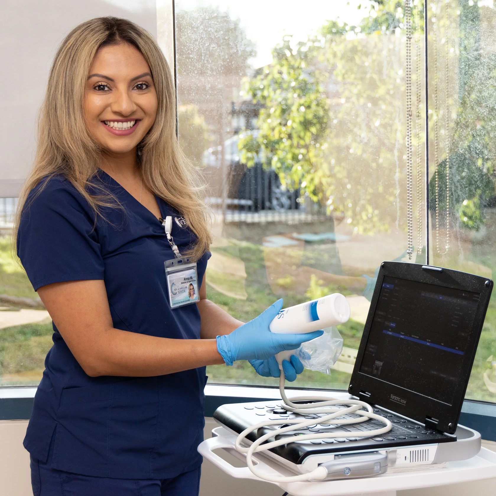
We offer convenient and coordinated care by offering our own on-site vascular laboratory. Our non-invasive vascular laboratory utilizes advanced imaging machines that look for vascular diseases that affect how well blood flows in the arteries and veins. We detect conditions not visible to the naked eye. Our laboratory is accredited by the Inter-societal Accreditation Commission (IAC) for Vascular Testing in veins.
Ultrasound Services are offered at both La Jolla and Vista locations! To make an ultrasound appointment in La Jolla, call 858-550-0330. To make an ultrasound appointment in Vista, call 760-249-7007
An ultrasound of the deep veins in your legs to rule out a blood clot
Time: 45 Minutes
No special preparation needed.
A venous insufficiency exam looks at the superficial veins in your legs to see if there is any reversed blood flow
Time: 75 Minutes
• Well hydrated • Do NOT wear compression stocking same day as appointment.
An upper extremity ultrasound of the veins in your arms to see if there are any blood clots
Time: 60 Minutes
No special preparation needed.
Ultrasound of the abdomen/pelvis post venous intervention of the iliac vein
Time: 45 Minutes
• Fasting 8-12 hours; • Water is OK; • Schedule in the morning.
What is vascular disease?
Vascular refers to the blood vessels in the body. There are two main types of blood vessels; the arteries and veins. Arteries bring oxygen-rich blood from the heart to every inch of the body; veins return the blood back to the heart and lungs for more oxygen. Vascular disease is when the blood vessels are no longer healthy and blood is not flowing efficiently.
Artery Disease
Carotid artery disease and stroke (TIA or Stroke)*
Lower extremity arterial disease (PAD)**
Upper extremity arterial disease
Abdominal aortic aneurysm (AAA)
Vein Disease
Varicose Veins
Chronic venous insufficiency
Deep Vein thrombosis
Vein Disease
Varicose Veins
Chronic venous insufficiency
Deep Vein thrombosis
What is Duplex Ultrasound
Duplex ultrasound involves using high frequency sound waves to look at the speed of blood flow, and structure of the blood vessels. The term “duplex” refers to the fact that two modes of ultrasound are used, Doppler and B-mode. The B-mode transducer obtains an image of the vessel being studied. The Doppler probe within the transducer evaluates the velocity and direction of blood flow in the vessel.
The Vascular Lab offers of:
Non-invasive testing
Same-day imaging appointments
Follow-up clinic visits with our on-site vascular team
Direct testing (duplex imaging)
Deep Vein Thrombosis- upper or lower extremity
Venous Reflux
Lower Extremity Duplex
book an appointment in Our center
To make an appointment or to inquire about how we can help you understand your treatment options.
The Duplex Ultrasound examination allows us to visualize the blood vessels that are not visible to the naked eye, even blood vessels that are deep within the muscles. Ultrasound looks at deep and superficial veins in the legs to check for venous-valvular incompetence (the underlying condition that causes varicose veins). The ultrasound examination is used to both identify the veins that have faulty valves and to map the anatomy of the veins, creating a ‘road map.’ This is necessary to make an accurate assessment of the cause and extent of the varicose veins, as well as to formulate the best treatment plan. This should be done for any individual being evaluated for varicose veins, leg swelling, patients who have failed prior treatment, patients who are symptomatic and in some patients with certain anatomic patterns of spider veins.
Before your test: This study does not require any preparation. You should not wear your compression stockings the same day as the examination. Make sure to be hydrated.
Vein Mapping
A vein mapping ultrasound is used to map the anatomy of the veins, creating a ‘road map.’ The vein diameters are also measured at various points. This is necessary to make an accurate assessment of the cause and extent of the varicose veins, as well as to formulate the best treatment plan. This study is performed prior to undergoing a vein procedure.
Before your test: This study does not require any preparation. You should not wear your compression stockings the same day as the examination. Make sure to be hydrated.
Deep Vein Thrombosis
A deep venous thrombosis (DVT) is a blood clot in the deep veins of the legs. This is a serious medical condition because the blood clot can travel from the deep venous system to the lungs and cause a lethal pulmonary embolism. A DVT can be diagnosed by an experienced vascular sonographer with a Duplex ultrasound examination. A DVT study is performed when a person has signs and symptoms of a lower extremity DVT such as leg pain and swelling. It may also be performed routinely after a vein procedure or other surgery.
Before your test: This study does not require any preparation.
Upper Extremity Venous Study
Ultrasound of the upper arms and neck. This study is performed to assess the deep and superficial venous system to determine the presence or absence when a person has signs and symptoms of a lower pain and swelling in arms.
Veins
Venous Reflux or Venous Insufficiency
The Duplex Ultrasound examination allows us to visualize the blood vessels that are not visible to the naked eye, even blood vessels that are deep within the muscles. Ultrasound looks at deep and superficial veins in the legs to check for venous-valvular incompetence (the underlying condition that causes varicose veins). The ultrasound examination is used to both identify the veins that have faulty valves and to map the anatomy of the veins, creating a ‘road map.’ This is necessary to make an accurate assessment of the cause and extent of the varicose veins, as well as to formulate the best treatment plan. This should be done for any individual being evaluated for varicose veins, leg swelling, patients who have failed prior treatment, patients who are symptomatic and in some patients with certain anatomic patterns of spider veins.
Before your test: This study does not require any preparation. You should not wear your compression stockings the same day as the examination. Make sure to be hydrated.
Vein Mapping
A vein mapping ultrasound is used to map the anatomy of the veins, creating a ‘road map.’ The vein diameters are also measured at various points. This is necessary to make an accurate assessment of the cause and extent of the varicose veins, as well as to formulate the best treatment plan. This study is performed prior to undergoing a vein procedure.
Before your test: This study does not require any preparation. You should not wear your compression stockings the same day as the examination. Make sure to be hydrated.
Deep Vein Thrombosis
A deep venous thrombosis (DVT) is a blood clot in the deep veins of the legs. This is a serious medical condition because the blood clot can travel from the deep venous system to the lungs and cause a lethal pulmonary embolism. A DVT can be diagnosed by an experienced vascular sonographer with a Duplex ultrasound examination. A DVT study is performed when a person has signs and symptoms of a lower extremity DVT such as leg pain and swelling. It may also be performed routinely after a vein procedure or other surgery.
Before your test: This study does not require any preparation.
Arteries
Arteries Testing and Screening
Who should get arterial screening?
In accordance with USPSTF and the AHA, ultrasound screening abdominal aortic aneurysm (AAA) with ultrasonography in men get 65-75 years who have ever smoked.
Because people with peripheral arterial disease (PAD) have this increased risk for heart attack and stroke, the American Heart Association encourages people at risk or over the age 55 to discuss PAD with their healthcare professional to ensure early diagnosis and treatment.
Upper Extremity Arterial Study (UEA)
Ultrasound of the arteries within the upper arms and neck. This study is designed to see if plaque has formed within the arteries, blocking the blood flow. This is also called atherosclerosis; hardening of the arteries.
Carotid Artery Ultrasound
Ultrasound of the carotid arteries in the neck to identify or rule out blockages or reduced blood flow from plaque build up that can lead to a stroke.
Abdominal Ultrasound
Ultrasound of the abdomen for identification and evaluation of aneurysmal disease of the aorta and its branches, such as an abdominal aortic aneurysm (AAA).
Lower Extremity Arterial study (LEA)
Ultrasound of the arteries within the lower legs. This study is designed to see if plaque has formed within the arteries, blocking the blood flow. This is also called atherosclerosis; hardening of the arteries.
Duplex Ultrasound
The duplex ultrasound evaluates the blood flow through your arteries and veins. There are several types of ultrasound tests.
Ankle-Brachial Index
The ankle-brachial index (ABI) is a test that uses inflatable pressure cuffs to assess blood flow in the legs and arms.
Carotid Artery Ultrasounds are routine evaluatons for symptoms of Transient Ischemic Attack (TIA) or stroke.
For more information on Vascular Health please visit our online library to view and download pdfs on many issues of vein disease.
Risk Factors for Stroke and Disease
High Blood Pressure
Tabacco Use
Diabetes
Family History
Age
Obesity
Sleep Apnea
Transient ischemic Attack (TIA)
Sudden Numbness
Trouble Speaking
Trouble Seeing
Dizziness
Severe Headache
Who Performs the test?
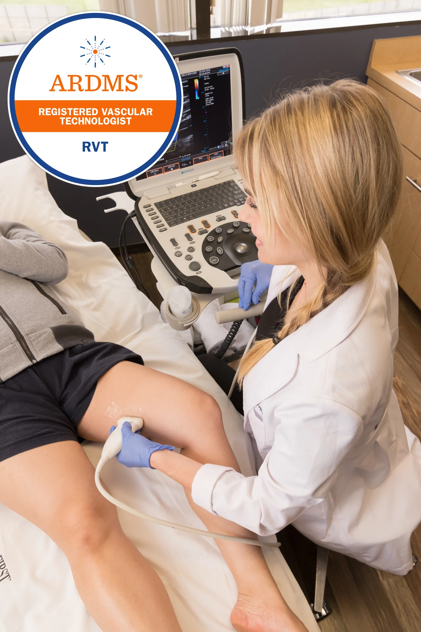
Accuracy is critical. Our ultrasounds are ALWAYS be performed by a credentialed sonographer, called a Registered Vascular Technologist (RVT). An RVT is a sonographer who completed a two-year ultrasound program, plus additional clinical training and obtained certification by meeting the highest standards by The American Registry for Diagnostic Medical Sonography® (ARDMS®). It is important that a specially trained RVT perform the study, because a special protocol must be followed for each study to meet accreditation standards. The protocol involves taking images at certain anatomic locations using special waveforms to show blood flow. All images taken by the RVT are reviewed by the physician.
Read What Our Patients Have to Say
What a wonderful experience, I love the site and the staff are super knowledgeable and helpful. To actually have someone assess and conduct an ultrasound on my legs and veins is an absolute dream come true. Thank you for your help!
Chelsey G.
Wow, where do I begin… how about with 10 stars (5 isn’t enough!). I am very impressed with La Jolla Vein & Vascular. I visited the Vista office and was greeted by the most friendly team ever. Everyone was so nice and welcoming, they really make you feel valued… that’s important especially when you deal with bad anxiety. But they will put you at ease and they know exactly what they are doing. Everything is thoughtfully explained and you feel confident in their diagnosis and treatment plan. The office is so clean and professional. I wish all medical offices, doctors and staff were like this!
Rebeca T.
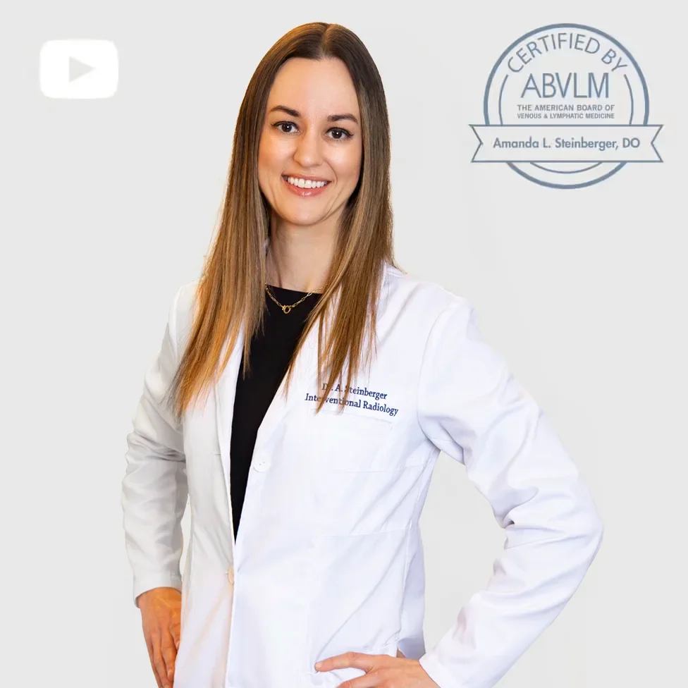
This practice is exceptional. All staff members are extremely nice and efficient. There is no wait time to be seen by appointment.Dr Steinberger is friendly, engaging, and highly competent. I am very satisfied with the treatment and the experience in general.
Samantha R.
For Health Care Professionals
How to Refer?
Fax a referral form with supporting documentation to 858-550-0676.
For same day appointments, check ‘STAT’ box on the order and call us at 858-550-0330.
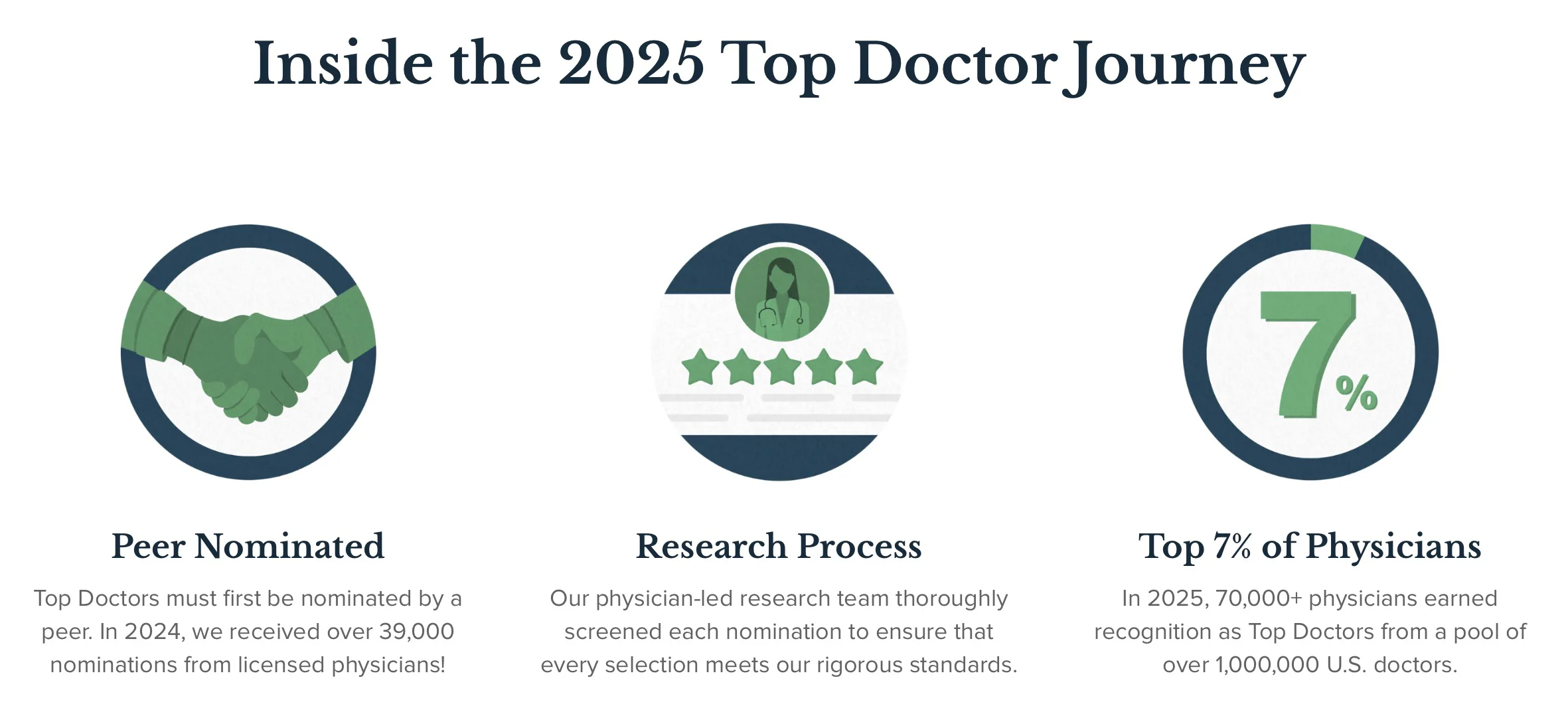
Castle Connolly Top Doctors represent the top 7% of all U.S. practicing physicians.
Castle Connolly Top Doctors are peer-nominated, and thoroughly vetted by our physician-led research team. These doctors are best-in-class healthcare providers, embodying excellence in clinical care as well as interpersonal skills.
Varithena® may be recommended based on your vein location, size, anatomy and vein tortuosity.
Varithena® is: • Effective to treat veins of different sizes above and below the knee • Requires no incisions, sedation or general anesthesia • Does not require a wire to be inserted along the length of your vein • Does not use heat, eliminating the risk of thermal injury
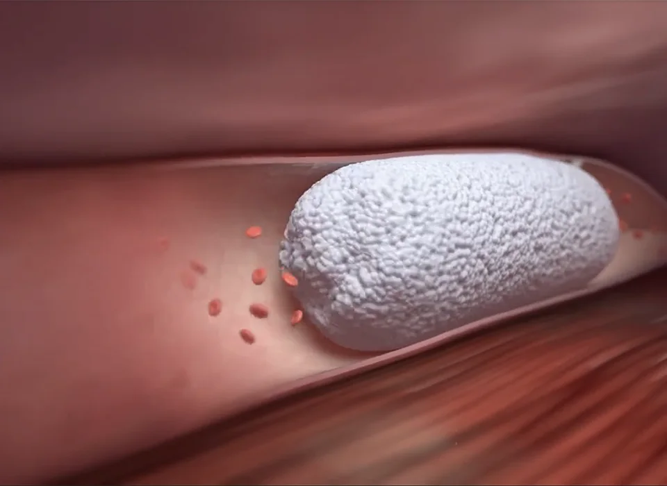
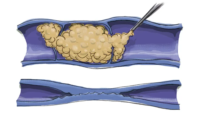
WHAT IS VARITHENA®?
Varithena® is a "microfoam" formulation of polidocanol (sclerosing agent) and CO2. The result is a microfoam that has an appearance and consistency similar to white, foamy shave cream. Ultrasound guidance is used to inject Varithena® microfoam into the vein. The microfoam fills the lumen for circumferential contact where it's designed to displace blood and destroy the endothelial lining efficiently.
Video below shows the proceedure.
How many treatments will I need?
The number and frequency of treatments depends on a patient's anatomy, how quickly the veins respond, and the patient's treatment goals. There is a limit on the amount of medication we can give you each day to avoid causing side effects. Some larger or resistant veins may require two treatments to respond completely but most veins respond to Varithena® after one treatment.
What should I expect on my treatment days?
You will sign your consent form then change into shorts provided by the office. We will clean your skin with alcohol, then use ultrasound to localize the veins. The foam medication will then be injected into your veins with a fine needle. The foam fills and treats the desired section of the vein. The diseased vein collapses and the foam is deactivated. Your legs will be elevated on a comfortable wedge pillow for approximately 15 minutes. After your treatment, we will help you into your compression stockings, then you will walk for 30 minutes prior to getting in your car.
Enter to receive special skinpen promotional offers!
Verificación de seguro gratuita. ¡Programe hoy mismo!
¿Puedo obtener cobertura de seguro para mi tratamiento de venas?
Dependiendo de su plan de seguro, es posible que tenga o no gastos de bolsillo. Trabajamos con sus aseguradoras para maximizar su cobertura y brindarle un resumen completo del costo de su tratamiento. Creemos en la transparencia de costos y le proporcionaremos un estimado de cualquier gasto de bolsillo previsto. El costo es importante. ¡Podemos colaborar con su seguro para obtener esta información hoy mismo!
Dr. Amanda Steinberger is a Harvard-trained, board-certified vascular interventional radiologist. She specializes in a broad range of superficial vein disease including varicose veins, spider veins, venous leg ulcers, lymphedema and cosmetic laser vein treatments. Dr. Steinberger sees patients in our La Jolla Office.
Free Insurance Verification. Schedule Today!
Can I get insurance coverage for my vein treatment?
Depending on your insurance plan, you may or may not have out of pocket expenses. We work with your insurance providers to maximize your insurance coverage and provide a complete overview of your treatment cost. We believe in cost transparency and will provide a cost estimate of any anticipated out of pocket expenses. Cost is important. We can work with your insurance to find out this information today!
To help make sure your payments are secured with the privacy you expect we have added a password protected payment page. Start by requesting a password be sent to your emall.
Can I get insurance coverage for my vein treatment?
With Molina insurance plans, most patients should not have out of pocket expenses. We work with Molina to maximize your insurance coverage and provide a complete overview of your treatment cost. We believe in cost transparency and will provide a cost estimate of any anticipated out of pocket expenses. Cost is important. We can work with your insurance to find out this information today!